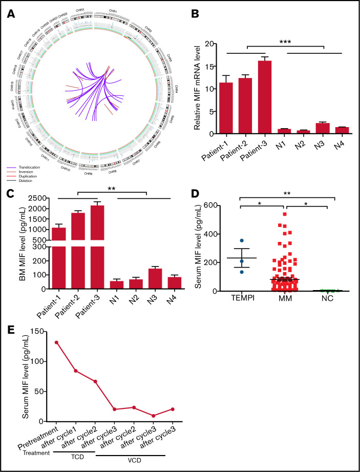
Figure 2.
Circos plot of somatic SVs and the expression of MIF in patients with TEMPI syndrome. (A) The center, color lines indicate the presence of translocations and inversions. In the middle ring, the red and black lines indicate presence of duplication and deletion, respectively. The outermost ring indicates chromosome number. (B-D) Expression level of MIF mRNA in CD138+ PCs (B), and secreted MIF protein in BM aspirates (C) from healthy individuals (N) and 3 patients with TEMPI syndrome. (D) The level of serum MIF in healthy individuals (NC) and patients with TEMPI syndrome and multiple myeloma (MM; n = 116). (E) Serum level of MIF in the patient 1 after treatment with TCD and VCD regimen. Statistical analyses were performed using the unpaired Student t test. *P < .05; **P < .01; ***P < .001.

