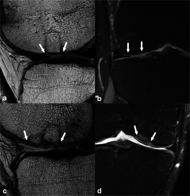
Fig. 2.
a, b A 31-year-old male patient 12 months after OAT with complete filling, a split-like integrational defect on the medial transplant border (depicted in image b), an intact surface, homogeneous structure, normal signal intensity of the repair tissue, no bony defect or overgrowth, and no subchondral changes, which resulted in an overall MOCART 2.0 knee score of 95 points. Also, the donor region can be appreciated in the anterior medial condyle in image a. c, d A 26-year-old male patient 9 years after OAT with an underfilling of 75–99% of the defect volume, complete integration, an irregular surface < 50% of repair tissue diameter, normal signal intensity, a bony defect ≥ transplant thickness, and no subchondral changes, which resulted in a MOCART 2.0 knee score of 75 points. Panels a and c are acquired with a sagittal proton-density-weighted turbo spin-echo sequence, whereas panels b and d are acquired with a coronal proton-density-weighted turbo spin-echo sequence with fat suppression. The white arrows indicate the repair tissue borders

