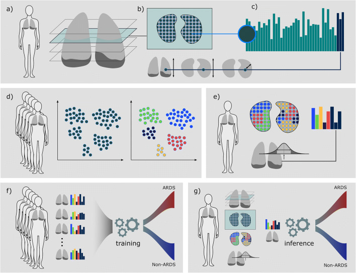
Fig. 1.
Overview—from CT scan to ARDS prediction: a We performed a machine learning–based segmentation of the lung, including effusion and air pockets. b From within the lung-mask, we extracted 2D radiomics features in a grid pattern over a kernel. In addition, we calculated the reference locations (anterior-posterior, inferior-superior, and the distance transform) to retrieve localized spatio-visual feature vectors for each location as illustrated in c. d A spatio-visual vocabulary sampled from feature vectors of the full dataset was learned. e After the vocabulary had been learned, a single patient is represented by his vocabulary histogram and statistical features calculated on the HU histogram of the full lung. f We trained a GBT ensemble on a training set of feature representations to distinguish cases that will develop ARDS in the future and cases that will not. g After training, prediction for a novel case is performed fully automated, from raw DICOM images, lung segmentation, and feature extraction to ARDS risk score. ARDS, acute respiratory distress syndrome; GBT, gradient boosted tree

