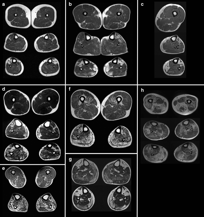
Fig. 2.
Magnetic resonance imaging (MRI) T1 sequences of patients with SMPX distal myopathy. a Patient F1 II.1 at 57 yrs showing normal thighs but fatty degeneration in lower legs: anterior compartment and medial gastrocnemius muscles. b F2 II.2 at 61 yrs showing minor degenerative change in thigh muscles semimembranosus, biceps femoris and left vastus intermedius; fatty degenerative changes in the anterior compartment muscles more on the left of proximal lower leg and fatty replacement of anterior compartment and part of the soleus muscles in the distal lower legs. c F3 II.1 at 58 yrs (left lower limb) with normal thigh and severe fatty degeneration in lower leg anterior compartment, medial gastrocnemius and medial part of distal soleus. d F6 II.1 at 52 yrs showing normal thigh and fatty degenerative changes in the anterior compartment (more on the right) and medial gastrocnemius muscles of proximal lower leg, and anterior compartment with soleus muscles in the distal lower legs. e F6 II.1 at 60 yrs with early degenerative changes in biceps femoris and semimembranosus in the thigh and more fatty degeneration of anterolateral compartments and of medial gastrocnemius and soleus in the lower legs. f F7 II.1 at 56 yrs shows milder fatty degeneration in vastus intermedius and medialis, semimembranosus and biceps femoris on the thigh, and in both posterior and anterior compartments on the lower legs, less in the lateral peroneal muscles. g F8 III.1 at 58 yrs shows lower legs with severe fatty replacement of anterior compartments and medial heads of gastrocnemius and milder changes in the soleus. h F9 II.1 at 76 yrs showing pronounced fatty degeneration in thighs: more in hamstrings than the lateral and intermediate vastus of the quadriceps, severe changes in distal lower leg anterior compartment and soleus muscles and peroneals more on the right

