Fig. 4.
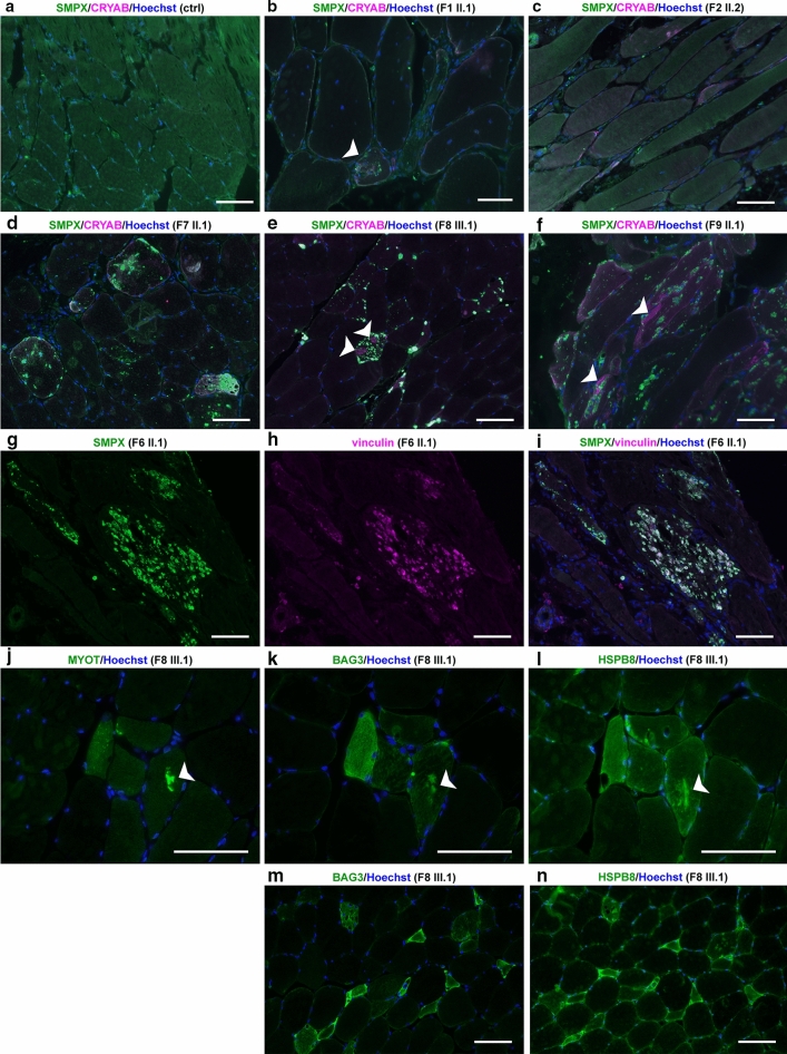
Immunofluorescence microscopy. a–f Immunofluorescent double staining; merged images are shown, SMPX is green and CRYAB is magenta. In control skeletal muscle (ctrl) free of neuromuscular disease, SMPX shows diffuse cytoplasmic and focal subsarcolemmal staining pattern. Muscle biopsy of F1 II.1 b shows moderate SMPX accumulation in a single fiber (white arrowhead), whereas in patient F2 II.2 c there is no SMPX accumulation. In probands from families F7, F8 and F9 (d–f), multiple SMPX-positive sarcoplasmic inclusions are present in several fibers. In F8 III.2 (e) and F9 II.1 (f), separate myofibrillar CRYAB accumulation is observed (white arrowheads), which does not co-localize with SMPX labeling. g–i Double immunostaining of F6 II.1. Single channel and merged images are shown; SMPX is green, vinculin is magenta. SMPX-positive protein inclusions (g) are also positive for vinculin (h), merged image (i). j–l; serial sections Myofibrillar accumulation (arrow) is positive for myotilin (j), and also for the CASA proteins BAG3 (k) and HSPB8 (l). m–n; serial sections) Several (atrophic) fibers show sarcoplasmic up-regulation of both BAG3 (m) and (n) HSPB8. Scale bars = 100 µm

