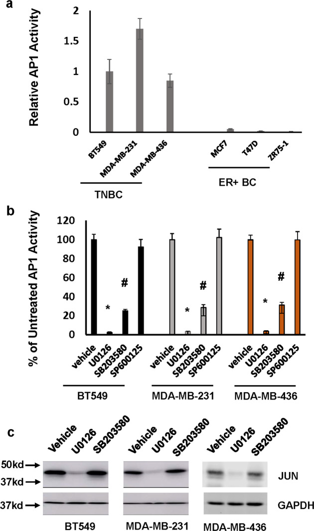
Fig. 1. Both ERK and p38MAPK signaling pathways contribute to sustained AP1 activity in TNBC cells.

a Endogenous AP1 activity in breast cancer cell lines analyzed by AP1 site-containing luciferase reporter plasmid. Data are means ± SD from three experiments. b BT549, MDA-MB-231, and MDA-MB-436 cells were treated with 5 µM U0126, SB203580, SP600125, or vehicle for 24 h followed by analyzing AP1 activity. Data are means ± SD from three experiments. *P < 0.001 vs vehicle; #P < 0.01 vs vehicle. c BT549, MDA-MB-231, and MDA-MB-436 cells were treated with 5 µM U0126, SB203580, or vehicle for 24 h followed by western analysis to detect JUN and GAPDH with the respective antibodies. Data are the representative of three independent experiments. All blots derived from the same experiment and were processed in parallel.
