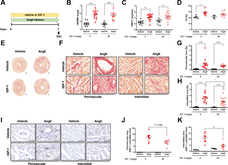
Fig. 4. Systemic infusion of low dose IGF-1 attenuates AngII-induced cardiac fibrosis.
A Schematic diagram of the experimental procedure. IGF-1 (20 ng/g/day) or vehicle was co-infused with AngII (3 µg/g/day) via a subcutaneously implanted osmotic minipump for 7 days in C57BL6 mice. B HW/BW ratio. Group sizes: vehicle (n = 10), AngII (n = 14), vehicle+IGF-1 (n = 10), AngII+IGF-1 (n = 14). C HW/TL ratio. Group sizes: vehicle (n = 10), AngII (n = 14), vehicle+IGF-1 (n = 10), AngII+IGF-1 (n = 14). D Echocardiographic analysis of fractional shortening (FS). Group sizes: vehicle (n = 5), AngII (n = 7), vehicle+IGF-1 (n = 4), AngII+IGF-1 (n = 7). E Representative Sirius red staining of hearts from AngII-infused mice treated with or without IGF-1. F Representative Sirius red staining of the perivascular and interstitial areas (magnification, x40, 20 µm). Quantification of perivascular fibrosis (G) and interstitial fibrosis (H). Group sizes: vehicle (n = 9), AngII (n = 13), vehicle+IGF-1 (n = 9), AngII+IGF-1 (n = 13). I Representative images of Ki67 immunostaining (magnification, ×40, 100 µm). Quantification of Ki67-positive perivascular cells (J) and interstitial cells (K). Group sizes: vehicle (n = 4), AngII (n = 7), vehicle+IGF-1 (n = 4), AngII+IGF-1 (n = 7). Data are presented as the mean ± SEM. *P < 0.05; **P < 0.01; ****P < 0.0001.

