Figure 4.
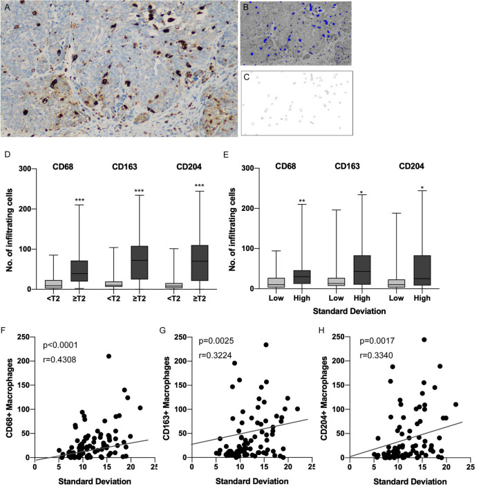
Tumors with heterogeneous internal texture in CT had more macrophage infiltration in UTUC. (A) Representative case of CD68 IHC that detected macrophages in muscle invasive urothelial carcinoma. (B) Image for detecting CD68-positive macrophages by the threshold using Image J software. Blue polygonal lesions indicate the CD68-positive macrophages. (C) Automatic counting of the number of infiltrating macrophages. (D) Boxplots for the number of infiltrating CD68-, CD163- and CD204-positive macrophages according to pathological T stage. (E) Boxplots for the number of infiltrating CD68-, CD163- and CD204-positive macrophages according to standard deviation obtained by TA. (F) Scatter plot comparing SD in TA and the number of CD68-positive macrophages. (G) Scatter plot comparing SD in TA and the number of CD163-positive macrophages. (H) Scatter plot comparing SD in TA and the number of CD204-positive macrophages. *p < 0.05, **p < 0.001, ***p < 0.0001. CT = computed tomography, UTUC = upper tract urothelial carcinoma, IHC = immunohistochemistry, TA = texture analysis.

