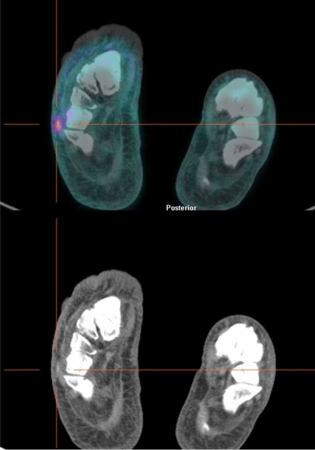
Fig. 1.

Axial views of [18F]FDG PET/CT images (top) and low-dose CT scan (bottom) of a diabetic patient with suspected osteomyelitis of right foot. The scan identified a focal uptake on a cutaneous/subcutaneous ulcer of the soft tissues of plantar surface without bone involvement, thus ruling out the diagnosis of osteomyelitis
