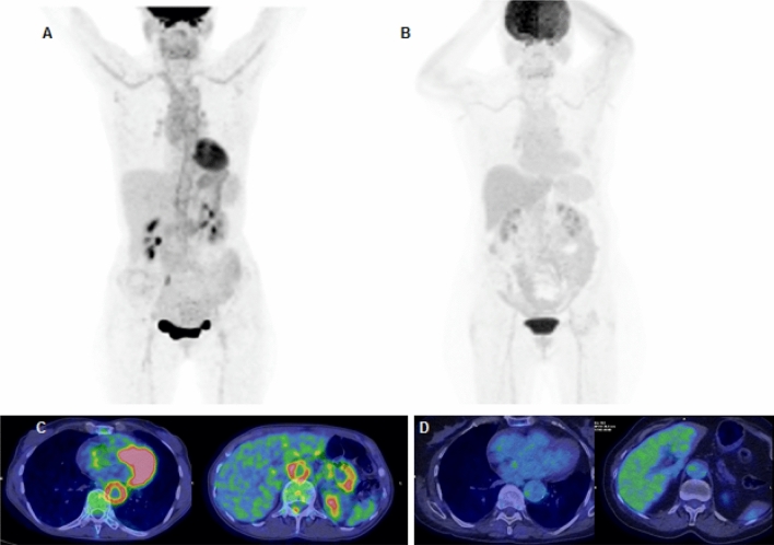
Fig. 2.
>50-year-old female with Giant Cell Arteritis (GCA). MIP (A, B) and axial PET/CT views before and after steroid treatment. Baseline (A, C): pathologic tracer uptake at ascending aorta, descending aorta, thoracic aorta, aortic arc, anonymous artery, subclavian arteries, common carotid arteries and abdominal aorta. Vascular tracer uptake greater than the hepatic one (grade 3 according to Meller reference scale). After treatment (B, D): no pathological uptake 3 months after steroid and immunosuppressive therapy (ongoing)

