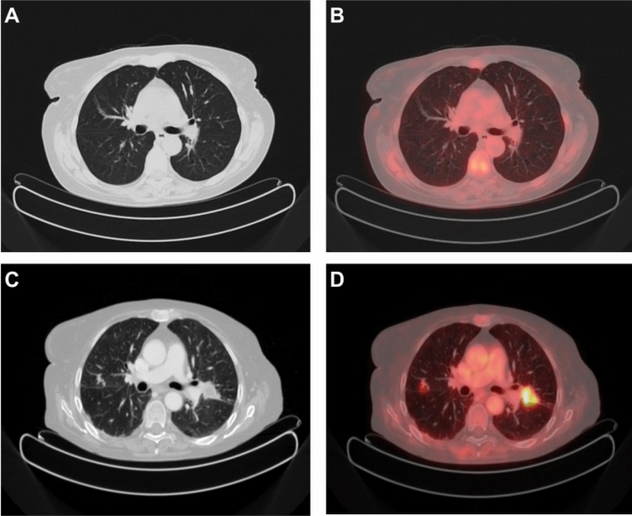
Fig. 5.
> 50-year-old woman with non-Hodgkin lymphoma, stage III, and secondary immunosuppression due to CHT and myelofibrosis. CT transaxial (A) and 18F-FDG PET/CT fused transaxial (B) imaging show mild pericardial and pleural effusion. The patient had fever without response to antibiotic therapy and pancytopenia. CT transaxial scan after contrast media injection (C) and 18F-FDG PET/CT fused transaxial (D) imaging after 6 months of corticosteroid therapy show persistent pericardial and pleural effusion and multiple pulmonary nodular lesions, later diagnosed as aspergillomas, which show intense 18F-FDG uptake. Typical Aspergillus lesion in left pulmonary hilar region, SUVmax 6.0

