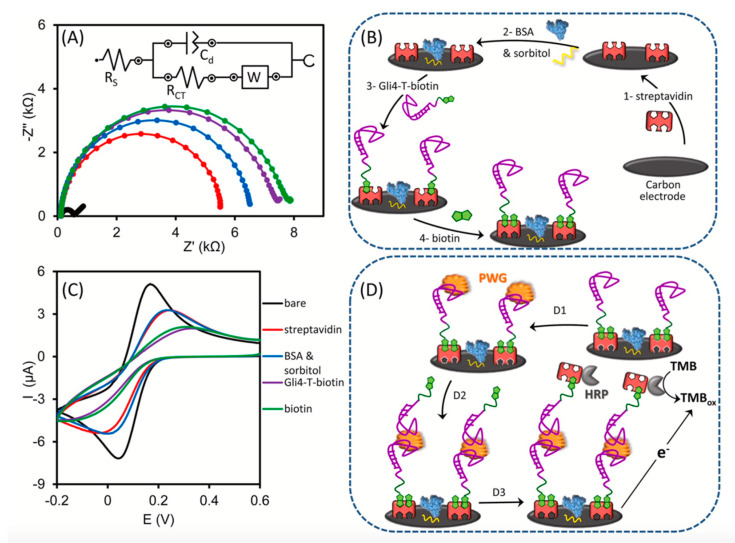
Figure 9.
Sandwich biosensor based on a truncated aptamer capable of recognizing gliadin directly in a DES: (A) Nyquist plots of 0.5 mM [Fe(CN)6]3−/4− after each modification step involved in the assembly of the biosensor, which are schematically represented in (B). (C) Voltammetric measurements recorded in KCl 0.1 M with 0.5 mM [Fe(CN)6]3− after each modification step. (D) Steps involved in the sandwich assay; D1: interaction with the sample containing gliadin, D2: incubation with the second aptamer labelled with biotin, D3: incubation with streptavidin–HRP conjugate and chronoamperometric measurement of the oxidized tetramethylbenzidine enzymatically produced. (Reprinted with permission from [69]).

