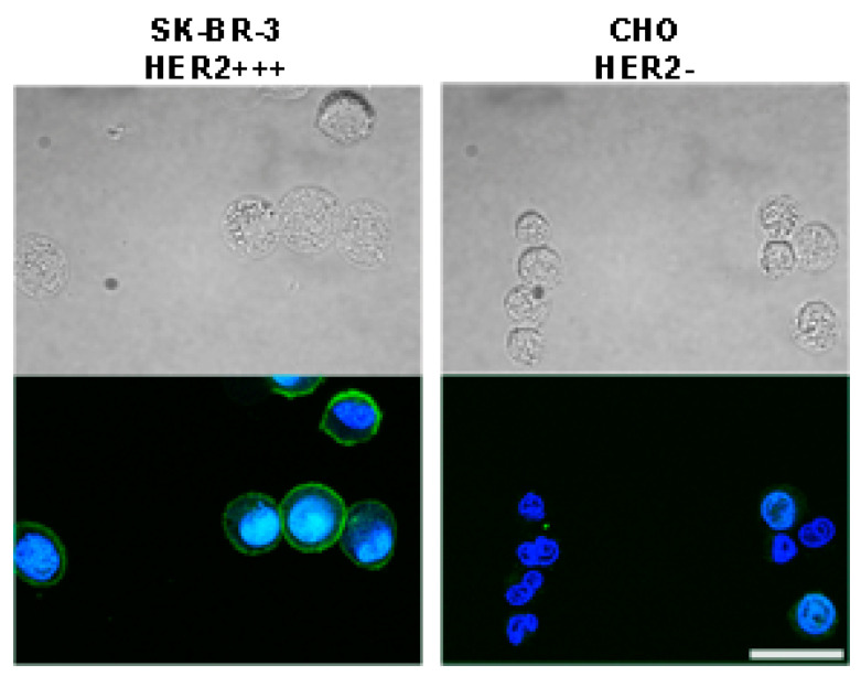
Figure 3.
Fluorescent immunostaining of cells possessing different HER2 expression levels with affibody ZHER2:342. Affibody ZHER2:342 was conjugated with FITC and cells were labeled with ZHER2:342-FITC. Top panels show bright-field images of SK-BR-3 and CHO cells, and bottom panels present overlaid confocal images of cells incubated with ZHER2:342-FITC and Hoechst 33342 (Hoechst 33342: excitation laser 405 nm, emission filter 445/45 nm; ZHER2:342-FITC: excitation laser 466 nm, emission filter 525/45 nm). Scale bar, 25 µm.

