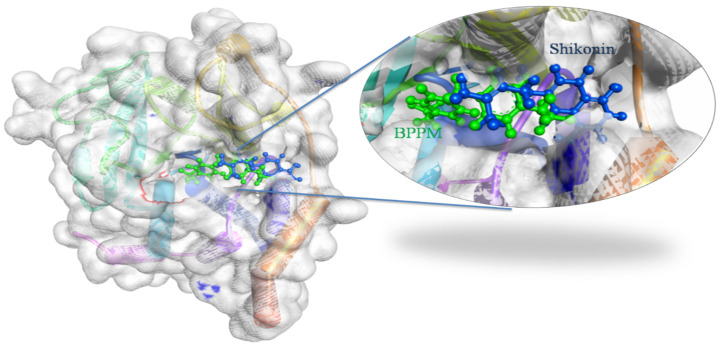
Figure 1.
Docking view of Shikonin in the binding sites of PTP1B (PDB ID: 1AAX). On the left side is the surface view of the docked complex-Shikonin and 1AAX, on the right side, a binding pocket is magnified to present Shikonin (shown in the blue-colored ball and stick view) superimposed on the co-crystal ligand-BPPM (shown in the green-colored ball and stick view). Diagrams are prepared in the Biovia Discovery Studio.

