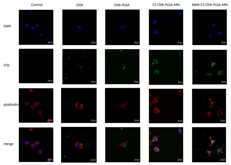
Figure 5.
Laser confocal scanning microscopy analysis of MAN-CS-OVA-PLGA-MP uptake by DCs. DCs were inoculated in a 6-well cell-culture plate on a round coverslip. After 24 h of culture, OVA-PLGA, CS-OVA-PLGA-MPs, MAN-CS-OVA-PLGA-MPs, or OVA was added separately. After an additional incubation for 12 h, the cells on the coverslips were fixed and stained using DAPI and Phalloidin-iFluor 555. Blue fluorescence indicates the nucleus, labeled by DAPI, while red fluorescence indicates the actin, stained with Phalloidin-iFluor 555. Cells were mounted with 90% glycerol and visualized using a confocal laser scanning microscope.

