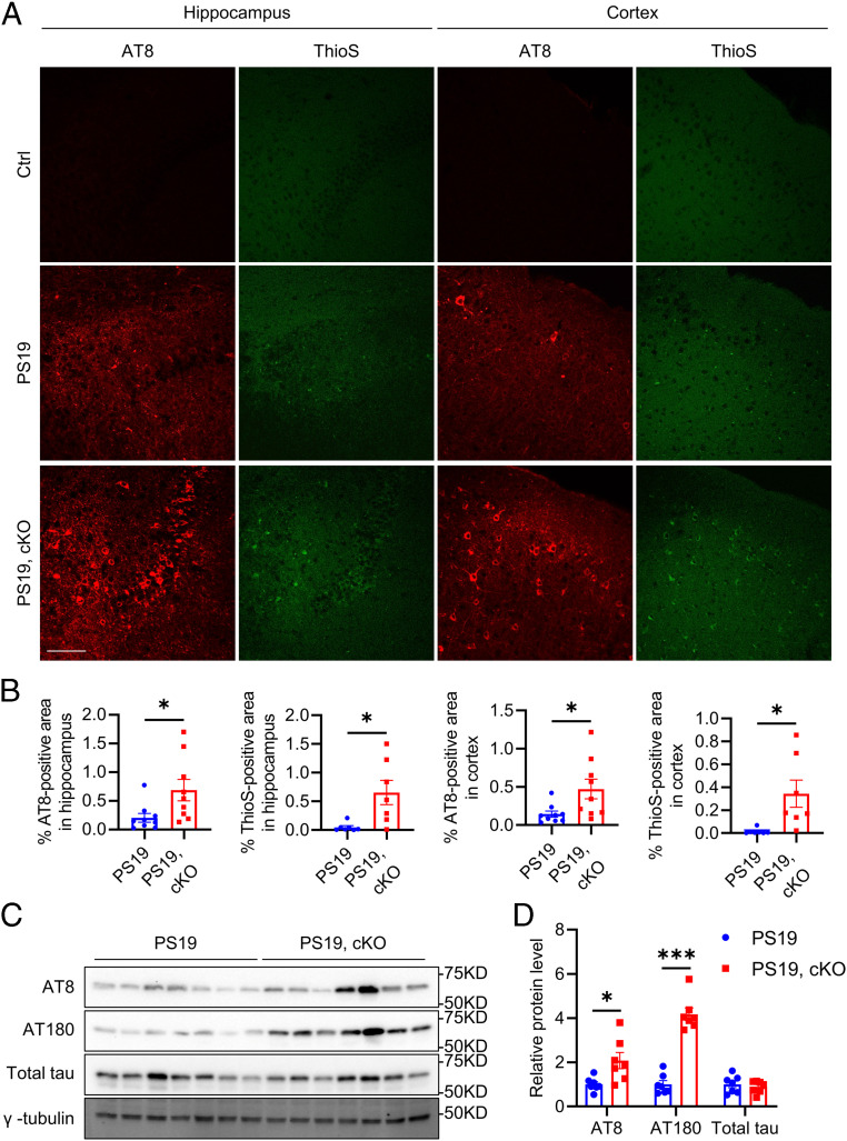
Fig. 6.
Microglial Atg7 cKO exacerbates tau pathology in vivo. (A) Representative immunofluorescence images of the hippocampus and cortex of control, PS19, and PS19, Atg7 cKO mice at 12 mo of age using the AT8 antibody and Thioflavin S dye. (Scale bar, 200 μm.) (B) Quantification of AT8-positive and ThioS-positive area in A. Two-tailed Student’s t test (AT8 staining: n = 9/group; ThioS staining: n = 7/group). (C) Representative Western blot image of phosphorylated tau (AT8, AT180) and total tau protein levels in brains of PS19 and PS19, Atg7 cKO mice at 12 mo of age. (D) Quantification of AT8, AT180, and total tau protein levels in C. Two-tailed Student’s t test (n = 7/group). Data are presented as mean ± SEM. *P ≤ 0.05; ***P ≤ 0.001.

