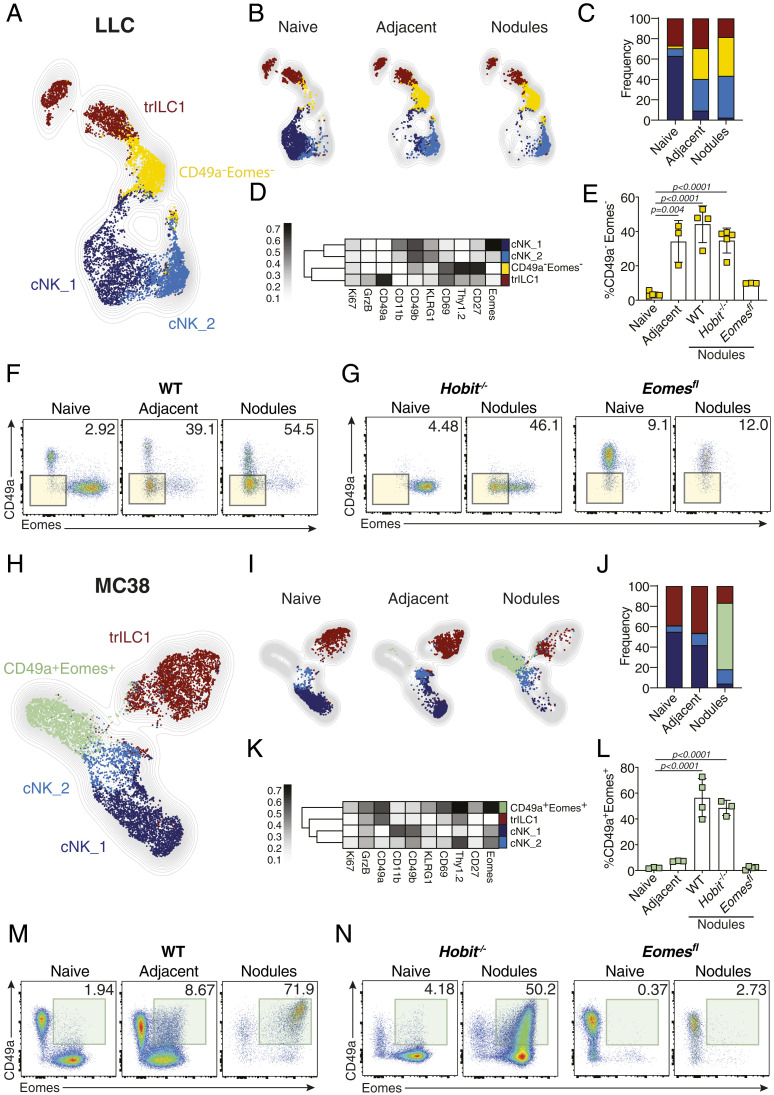
Fig. 4.
Metastatic livers drive the differentiation of unique cNK populations. NKp46+ cells from naïve and day 21 metastatic (nodules and adjacent tissue) livers were analyzed by multiparameter single-cell mapping using flow cytometry. (A–G) LLC metastatic and control livers. (H–N) MC38 metastatic and control livers. Samples were pregated on single live CD45+lineage− cells and subsequently gated on NK1.1+NKp46+ cells. cNK = conventional NK cells, CD49a−CD49b+; trILC1 = tissue-resident ILC1s, CD49a+CD49b−. (A and H) UMAP projections overlaid with FlowSOM-guided manual metaclusters displaying cNKs and trILC1s from all samples. (B and I) UMAP projections overlaid with FlowSOM-guided manual metaclusters separated by sample category (naïve, adjacent, nodules). (C and J) Relative frequency of each cluster in the different sample categories (naïve, adjacent, nodules). (D and K) Heat map summary of median marker expression values of the different markers analyzed for each cluster. (E and L) The frequency of unique metastasis-induced subsets. (E) CD49a−Eomes− NKp46+ cells in LLC metastasis. (L) CD49a+Eomes+ NKp46+ cells in MC38 metastasis. The bar represents the mean ± SD, and the symbols represent livers from individual mice. One-way ANOVA with Tukey’s multiple comparisons test. Experiments were performed at least twice with similar results. (F and M) Representative dot plots of cNKs and trLC1s cells for each sample category based on their expression of CD49a and Eomes. (F) LLC metastasis. Highlighted in yellow is the CD49a−Eomes− population observed in LLC-adjacent tissue and nodules. (M) MC38 metastasis. Highlighted in green is the CD49a+Eomes+ population observed in MC38 nodules. (G and N) Representative dot plots of cNKs and trILC1s isolated from naïve liver or metastatic nodules from Hobit−/− and Eomesfl mice based on their expression of CD49a and Eomes. (G) LLC metastasis. Highlighted in yellow is the CD49a−Eomes− population observed in LLC-adjacent tissue and nodules. (N) MC38 metastasis. Highlighted in green is the CD49a+Eomes+ population observed in MC38 nodules. Groups contained three to five mice. Experiments were performed at least twice with similar results.

