Fig. 4.
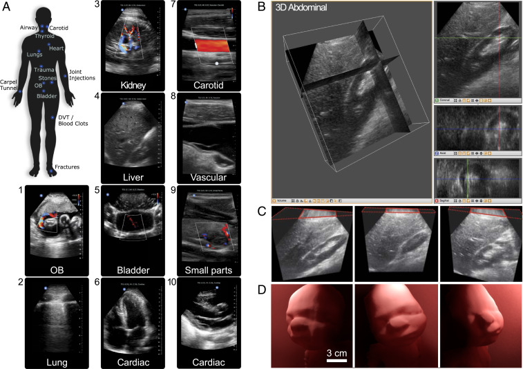
Ultrasound imaging with the UoC. (A) Labeled areas of the whole body where the UoC probe enables imaging. Example ultrasound imaging from several presets are labeled, including (1) OB: fetus umbilical cord Doppler flow; (2) lung: pleura interface; (3) abdominal: kidney vascular flow; (4) abdominal: liver; (5) pelvis: bladder jets; (6) cardiac: apical four chamber; (7) vascular: carotid artery; (8) vascular: internal jugular vein; (9) small organ: thyroid nodule; (10) cardiac: parasternal long axis. (B) Three-dimensional ultrasound renders of an in vivo human abdomen scan volume, 15 cm deep and 12 cm × 12 cm wide, where the structures and orientation between the kidney and liver can be viewed in multiple slices. (Left) Multiplanar reconstruction visualized in 3D with orthogonal slices and (Right) individual 2D image slices for coronal (XZ), axial (XY), and sagittal (YZ). (C, Left) Slice showing length of kidney and details of the renal cortex. (C, Middle) Slice showing the liver and edge of the kidney (Morison’s pouch). (C, Right) Slice showing liver parenchyma and kidney parenchyma. (D) Three-dimensional ultrasound of a 36-wk-old fetal ultrasound training phantom where surface rendering on a computer highlights contours of the baby’s face from different view angles. The ultrasound scan volume is 15 cm deep and 12 cm × 12 cm wide.

