Fig. 3.
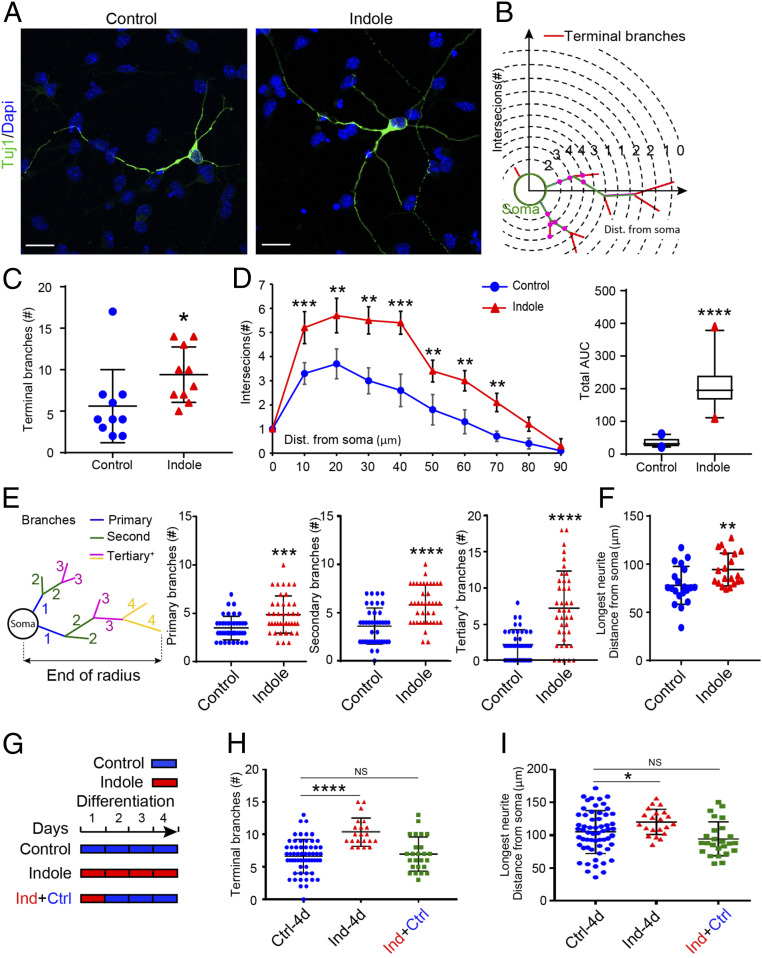
Indole promotes neuronal maturation ex vivo. (A) Representative images of Tuj1-immunostained neurons (green) in NPC cultures treated with indole- (100 μM) or vehicle-supplemented media for 4 d. Nuclei (blue) stained with DAPI. (Scale bar: 20 μm.) (B) Diagram indicating the quantification of neurite branching by Sholl analysis. (C and D) Quantification of total number of terminal branches revealed a significant effect of indole to promote neuronal maturation (D, Left) Sholl analysis quantification of intersections with distance from the cell soma. (Right) The corresponding total AUC in the Sholl plot. (E) Diagram indicating the quantification of neurite branching by I/O scheme analysis and quantification of neurite branches showing significant increase in primary, secondary, and tertiary in indole-treated NPCs. (F) Quantification of longest neurite length revealed a significant increase in neurons differentiated in indole-treated media. (G) Timeline for treatment of NPCs with indole (100 μM) at different time points during neuronal differentiation. (H and I) Quantification of terminal branch number (H) and longest neurite length (I) revealed that the differentiation of NPCs in indole-supplemented media for 24 h followed by 3 d with vehicle media failed to enhance neuronal maturation (n > 10 neurons per coverslip [each circle or triangle for one neuron] from n = 3 cultures per treatment condition). Data are presented as mean ± SEM with the exception of D, Right in which the horizontal line in the box plot represents the mean, and the whiskers show the 10 to 90th percentile. Statistical differences were determined using Mann–Whitney U test (C–F), Kruskal–Wallis test (H), and one-way ANOVA (I). Asterisks indicate a significant difference between groups (****P < 0.0001, ***P < 0.001, **P < 0.01, *P < 0.05, and NS represents nonsignificant differences).

