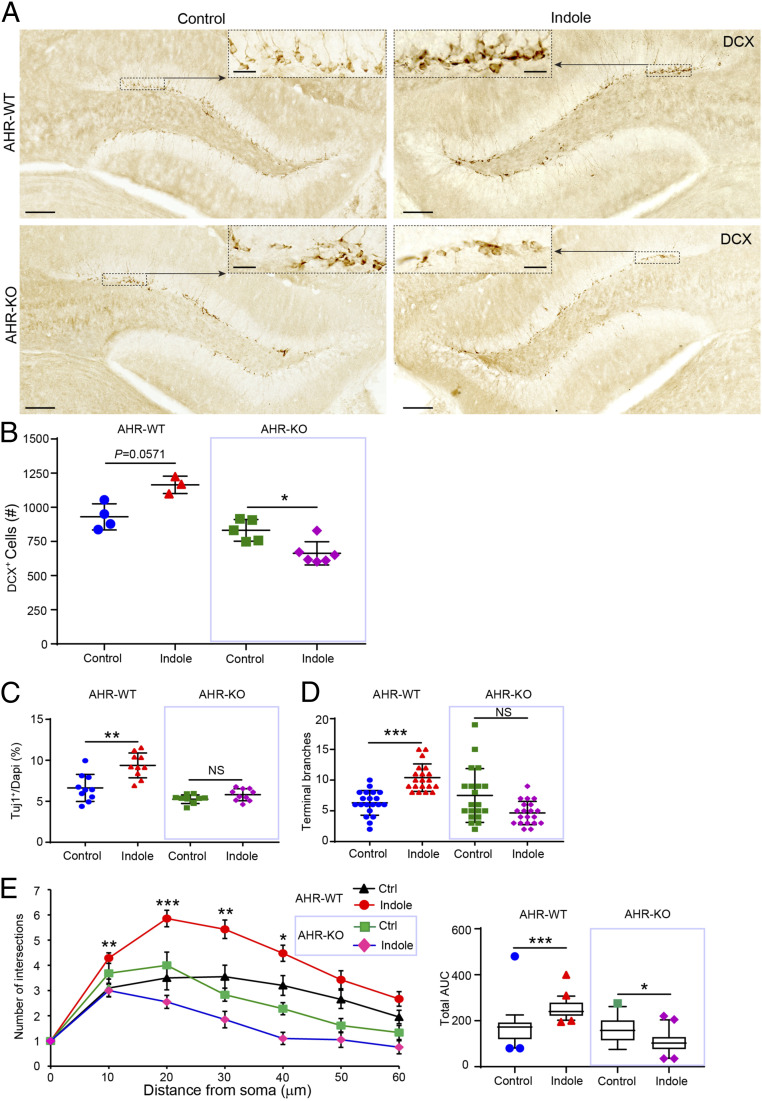
Fig. 5.
Indole promotes neurogenesis via the AhR ex vivo and in vivo. (A) Representative images of DCX-DAB–stained immature neurons in the DGs of SPF mice fed standard or indole-supplemented water (200 μM) for 5 wk. The black dashed boxes indicate comparative areas that are magnified to show the notable increase in DCX+ neurons in indole-supplemented AhR-WT but not AhR-KO mouse dentate gyri. (B) Quantification of DCX+ immature neuron populations in the DGs of vehicle-treated AhR-WT mice (n = 4) compared with indole-treated AhR-WT mice (n = 3) and vehicle-treated AhR-KO mice (n = 5) compared with indole-treated AhR-KO mice (n = 6). (C) Quantification of Tuj1+ neurons revealed indole enhances neurogenesis in AhR-WT NPCs but not AhR-KO NPCs. (D and E) Quantification of neurite branching revealed a significant effect of indole to promote neuronal maturation in AhR-WT NPCs but not AhR-KO NPCs. (E, Left) Sholl analysis quantification of intersections with distance from the cell soma (n > 10 neurons per coverslip [each circle or triangle for one neuron] from n = 3 cultures per treatment condition). (Right) The corresponding total AUC in the Sholl plot. Data are presented as mean ± 2 SEM with the exception of E, Right in which the horizontal line in the box plot represents the mean and the whiskers show the 10 to 90th percentile. Statistical differences were determined using the Kruskal–Wallis test for nonparametric data. Asterisks indicate a significant difference between groups (***P < 0.001, **P < 0.01, *P < 0.05, and NS represents nonsignificant differences).

