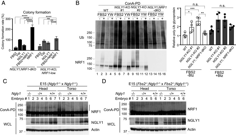
Fig. 5.
NRF1 is primarily responsible for the induction of cytotoxicity in NGLY1-KO cells. (A) Colony formation assays in NGLY1;NRF1-dKO and NGLY1-KO;NRF1-low HeLa cells overexpressing the indicated proteins. Colony formation rates were calculated relative to the number of colonies in GFP-overexpressing cells. Cells were counted in each of four replicates. (B) Accumulation of ubiquitinated glycoproteins in wild-type (WT), NGLY1-KO, and NGLY1;NRF1-dKO HeLa cells overexpressing FBS2. (Left) Ubiquitination of glycoproteins concentrated with concanavalin A (ConA)–conjugated agarose were analyzed by immunoblotting with anti-ubiquitin (Ub) antibody. (Right) Ubiquitinated glycoprotein levels (average intensities between 100 and 250 kDa) were normalized against the level of glycoprotein ubiquitination in FBS2 YW/AA–overexpressing WT cells. Error bars show means ± SD from four biological replicates. (C and D) Ubiquitination of NRF1 in Ngly1-KO (C) or Fbs2;Ngly1 dKO (D) day 15 embryos. ConA-bound glycoproteins from whole-cell lysates (WCL) were subjected to immunoblotting with anti-NRF1 antibody. Vertical line: ubiquitinated NRF1. Statistical significance was determined by one-way ANOVA with Dunnett’s posttest (A) or one-way ANOVA with Tukey’s posttest (B). ****P < 0.0001; ***P < 0.0002; **P < 0.002; *P < 0.03, n.s., not significant.

