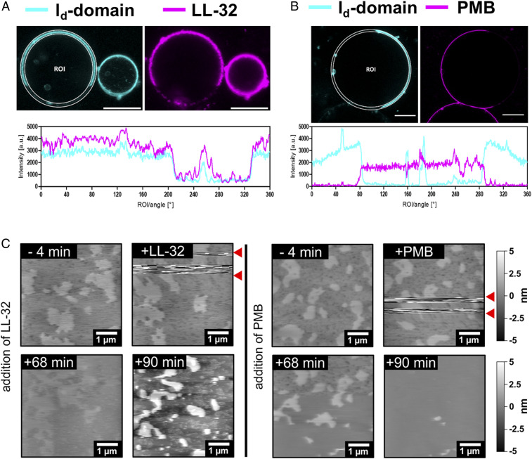
Fig. 5.
LL-32 and PMB interact via opposing interaction sites on cholesterol-rich model membranes. (A and B) False-color presentation of (A) LL-32-Rho and (B) PMB-BODIPY on giant vesicles reconstituted from DOPC:SM:Chol (2:2:1 M). The ld domain (cyan) of the membrane was labeled using the lipid-dye conjugate PC-BODIPY or Atto633-DOPE and cholesterol-rich (lo-) domains appear black. Peptides were added at 4.5 μM LL-32-Rho or 22.5 μM PMB-BODIPY (magenta). (Scale bars, 10 μm.) The data are representative of n = 3 independent experiments. (C) AFM images of solid-supported bilayers of the macrophage mimetic lipid mixture PLMAK:SM:Chol (2:0.5:0.2 M). Bilayers were immobilized on mica and peptides were added to a final concentration of 25 µM. The peptides were added in between the two red arrows and were responsible for the visible disturbances. All AFM images were only flattened and not further processed. The presented images were obtained before and after the addition of peptide or buffer at the indicated times. The data are representative of n = 2 (LL-32) and n = 6 (PMB) independent experiments.

