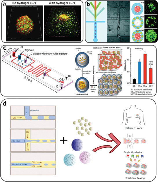
Figure 2.

a) Fluorescence microscopy images of MCTS co‐cultures of fibroblasts (green) and cancer cells (red). Tumor invasion occurs only by MCTSs surrounded by the collagen‐alginate 3D hydrogel. Reproduced with permission.[ 33 ] Copyright 2018, Elsevier. b) Microfluidic assisted formation of liver‐in‐a‐droplet. Microfluidic production of liver‐in‐a‐droplet, where hepatocytes and fibroblasts were encapsulated in the core and shell of the capsule, respectively. Reproduced with permission.[ 62 ] Copyright 2016, Royal Society of Chemistry. c) A microfluidics device used for production of collagen core and alginate shell capsules used for 3D tumor vascularization experiments. 3D vascularized tumors expressed increased drug resistance in the presence of a commonly used chemotherapeutic drug, although this resistance reduced when treated with drug carrying nanoparticles. Reproduced with permission.[ 63 ] Copyright 2017, American Chemical Society. d) The rationale of combining microfluidics, hydrogels and cancer cell encapsulation in order to produce high throughput personalized drug treatments. Reproduced with permission.[ 46 ] Copyright 2019, John Wiley and Sons.[ 64 ] Copyright 2020, American Chemical Society.
