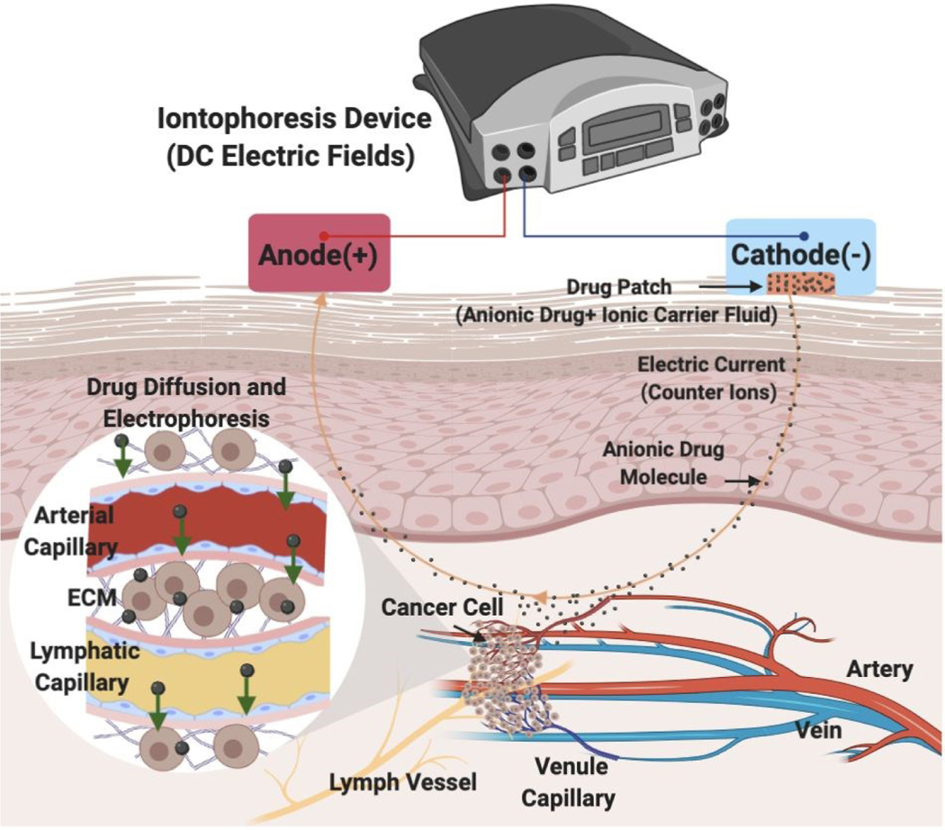
Figure 1. Iontophoretic transdermal drug delivery into the tumor vasculature in vivo.
The schematic shows the in vivo transport of an anionic drug from the artery to the tumor where it eventually drains into the lymph vessel. Iontophoretic transdermal drug delivery includes insertion of the cathode along with the drug patch on the skin from where the anionic drug molecules move to the tumor facilitated by the transport of the counter ions (from carrier solvent) to the anode.

