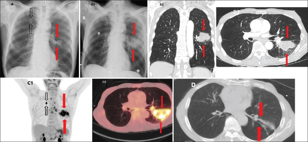
Figure 1.
Computed tomography and fluorodeoxyglucose positron emission tomography modality imaging of cocaine-inducible focal alveolar hemorrhage reveals rapid doubling and resolution and exhibits a solid-like, hazy appearance and preserved underlying bronchial structures and vasculature. (A) Initial chest X-ray showing two lung nodules up to 2.7 cm (red arrows – left lower lobe, black arrows – right upper lobe. (B1) Subsequent chest X-ray illustrating left lower lobe nodule increase to 5 cm (red arrows). (B2) Subsequent coronal chest computed tomography illustrating left lower lobe nodule increase to 5 cm (red arrows). (B3) Subsequent axial chest computed tomography illustrating left lower lobe nodule increase to 5 cm (red arrows). (C1) Intensely avid, 7 cm lower lobe nodule lesion on coronal fluorodeoxyglucose positron emission tomography/computed tomography with photopenic areas suggestive of necrosis (red arrows) with additional intensely avid right upper lobe nodule (black arrows). (C2) Intensely avid, 7 cm lobe nodule lesion on axial fluorodeoxyglucose positron emission tomography/computed tomography with photopenic areas suggestive of necrosis (red arrows). (D) Resolved pulmonary lesion with minimal residual fibrosis on axial chest computed tomography 1 month after initial imaging (red arrows)

