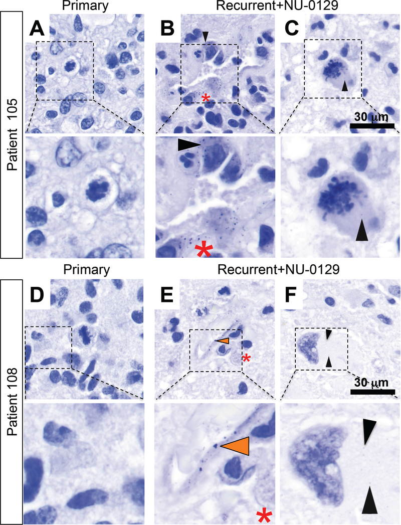
Fig. 5. Silver staining of pre- and post-treatment tumors 105 and 108.
(A, D) Silver staining of pre-treatment tumors in patients 105 and 108. (B-C; E-F) Silver staining of tumor sections post-NU-0129 treatment. In tumors resected after NU-0129 administration, Au was present within endothelial cells (E, orange arrowhead), tumor cells (B, C, F; black arrow head), and macrophages (B, E, red asterisk). Cell shown in panel F demonstrates therapy-related nuclear atypia typical for a post-therapy GBM tumor cell (panel F).

