Fig. 1.

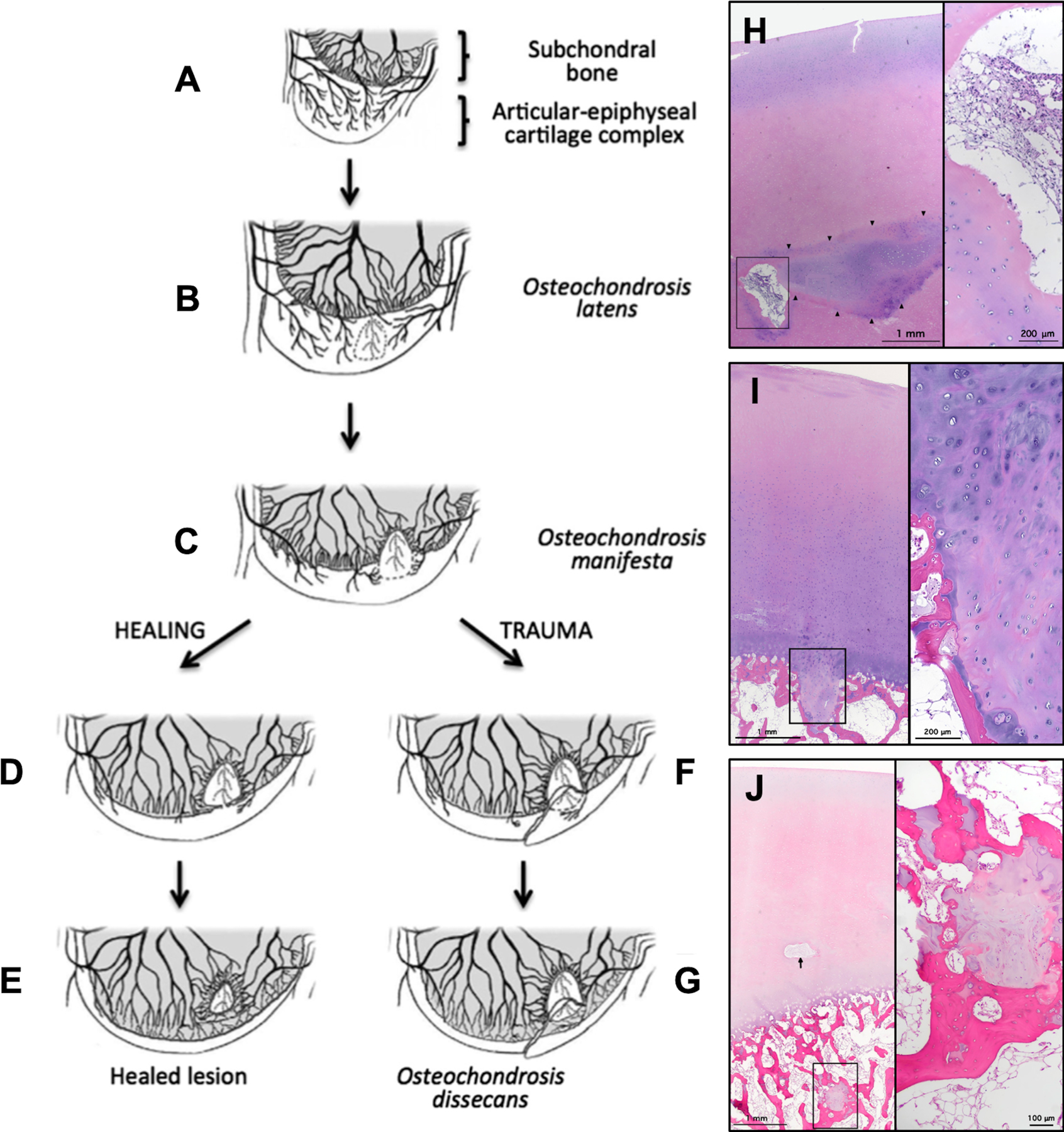
Figs. 1-A through 1-J Diagrams and photomicrographs demonstrating the pathogenesis of osteochondrosis in animals (Figs. 1-A through 1-G) and OCD in human pediatric cadavers (Figs. 1-H, 1-I, and 1-J). (Figures 1-A through 1-G modified, with permission, from: McCoy AM, Toth F, Dolvik NI, Ekman S, Ellermann J, Olstad K, Ytrehus B, Carlson CS. Articular osteochondrosis: a comparison of naturally-occurring human and animal disease. Osteoarthritis Cartilage. 2013 Nov;21[11]:1638–47; Ytrehus B, Carlson CS, Ekman S. Etiology and Pathogenesis of Osteochondrosis. Vet Pathol. 2007 Jul;44[4]:429–48; and Ekman S, Carlson C. The pathophysiology of osteochondrosis. Vet Clin North Am Small Anim Pract. 1998 Jan;28[1]:17–32; and Figures 1-H, 1-I, and 1-J modified from: Tóth F, Tompkins MA, Shea KG, Ellermann JM, Carlson CS. Identification of areas of epiphyseal cartilage necrosis at predilection sites of juvenile osteochondritis dissecans in pediatric cadavers. J Bone Joint Surg Am. 2018 Dec 19;100[24]:2132–39.) Fig. 1-A Normal endochondral ossification. Fig. 1-B Development of osteochondrosis latens because of failure of the cartilage canal blood supply, resulting in epiphyseal cartilage necrosis. Fig. 1-C Osteochondrosis manifesta lesion develops as a delay in the progression of the ossification front. Figs. 1-D and 1-E Healing of an osteochondrosis manifesta lesion by incorporation into subchondral bone. Figs. 1-F and 1-G Development of an OCD lesion as a result of trauma, causing collapse of articular cartilage overlying areas of necrotic epiphyseal cartilage. Fig. 1-H Hematoxylin and eosin staining of articular and epiphyseal cartilage from the central aspect of the medial femoral condyle from a 4-year-old female donor containing an osteochondrosis latens lesion. Arrowheads demarcate necrotic epiphyseal cartilage. Right: High-magnification image demonstrates cartilage canal containing multiple degenerate vessels. Fig. 1-I Hematoxylin and eosin staining of articular and epiphyseal cartilage from the central aspect of the medial femoral condyle from a 9-year-old male donor containing an osteochondrosis manifesta lesion. Right: High-magnification image demonstrates necrotic epiphyseal cartilage partially surrounded by bone. Fig. 1-J Hematoxylin and eosin staining of articular and epiphyseal cartilage from the central aspect of the lateral femoral condyle from a 4-year-old female donor containing a healing osteochondrosis manifesta lesion. Arrow points to a degenerate cartilage canal. Right: High-magnification image demonstrates necrotic epiphyseal cartilage completely surrounded by bone.
