Fig. 8.

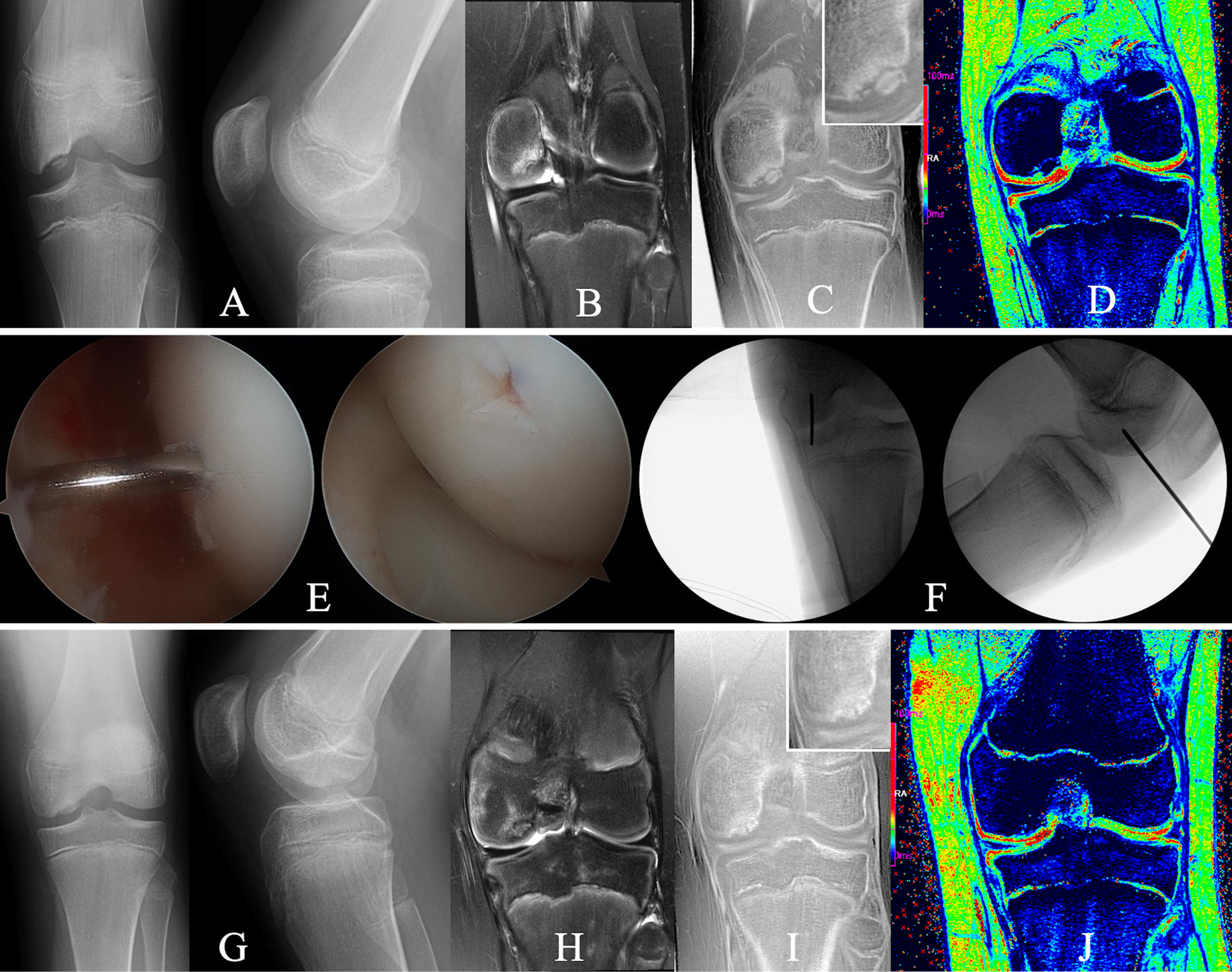
Figs. 8-A through 8-J An 11-year-old boy who underwent transarticular drilling for treatment of an OCD lesion in the medial femoral condyle of the left knee. Fig. 8-A Preoperative tunnel and lateral radiographs reveal an osseus OCD lesion. Figs. 8-B, 8-C, and 8-D Preoperative images made with advanced clinical 3-T MRI protocol, obtained with same imaging parameters as in Figure 3, including a T2-weighted coronal image demonstrating marrow edema (Fig. 8-B), a CT-like image contrast bone window (Fig. 8-C) depicting a partially ossified progeny lesion without evidence of osseous bridging with the parent bone (insert shows magnification of the lesion), and T2* map (Fig. 8-D), which provides additional information, specifically on cartilage integrity showing intact residual epiphyseal cartilage (green) and articular cartilage (red). Fig. 8-E Arthroscopic photographs made during transarticular drilling with a 0.045-in (0.114-cm) Kirschner wire to penetrate the lesion in a perpendicular fashion through the articular cartilage. Fig. 8-F Fluoroscopy confirming wire placement. Fig. 8-G Radiographs made 3 months postoperatively demonstrating interval healing. Figs. 8-H, 8-I, and 8-J Comprehensive MRI assessment demonstrating mild residual edema in parent bone and lesion in T2-weighted coronal MRI images (Fig. 8-H); solid osseous bridging and further ossification throughout the entire lesion in the CT-like contrast bone window images (insert shows magnification of the lesion) (Fig. 8-I), which was demonstrated on all slices (not shown); and preserved articular cartilage (red) after surgical intervention (see T2* map), which is mostly for further prognostication (Fig. 8-J). The patient returned to all activities.
