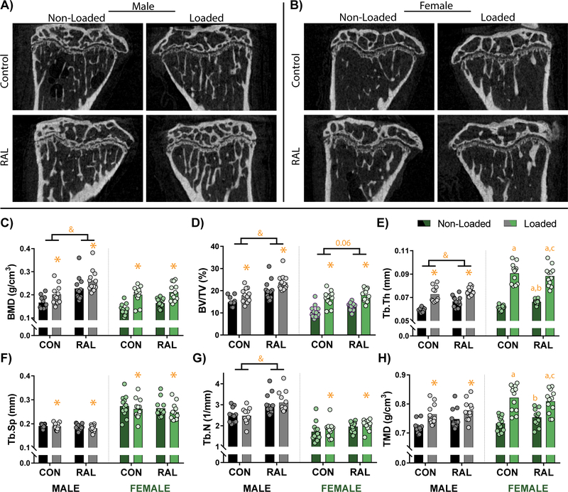
Figure 1.
Sagittal CT images of the proximal tibia from (A) male and (B) female mice qualitatively show increased cancellous bone due to loading and RAL. (C) Quantitative measures of bone mineral density (BMD) and (D) bone volume fraction (BV/TV) similarly indicate additive mass-based effects due to loading and RAL in males. In females, the loading had the more prominent effect than RAL. (E) These improvements were driven by load- and RAL-based increases to trabecular thickness (Tb.Th), (F) load-based decreases to trabecular spacing (Tb.Sp), and (G) load- and RAL-based increases in trabecular number (Tb.N). (H) In addition, tissue mineral density (TMD) was increased due to loading in both males and females. For a two-way ANOVA, a ‘&’ indicates a main effect of RAL and an ‘*’ indicates a main effect of loading. If the interaction term was significant, an ‘a’ indicates a significant difference from non-loaded control, ‘b’ indicates a significant difference from loaded control, and ‘c’ indicates a significant difference from non-loaded RAL.

