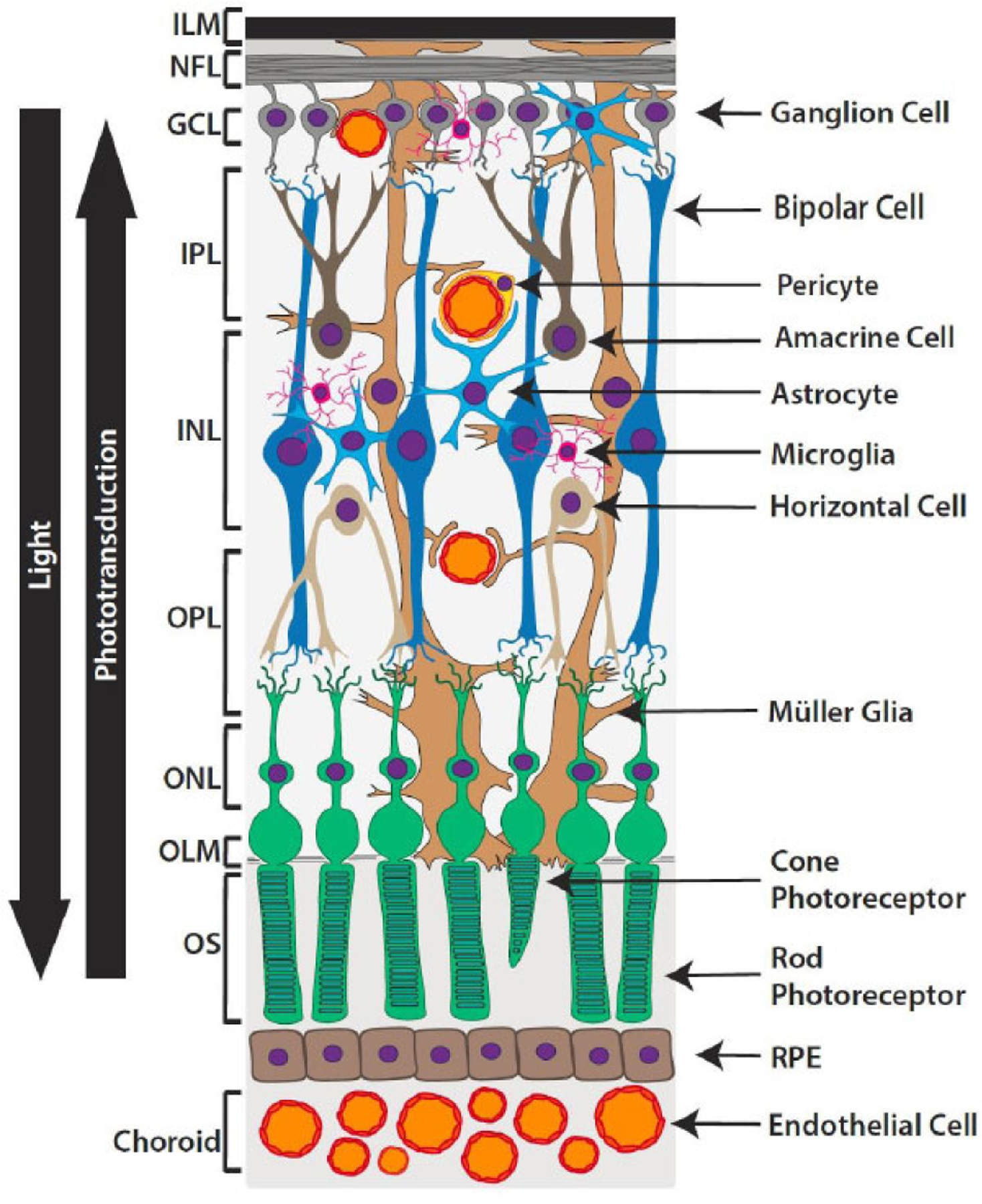
Fig. (1). Diagram of the retinal neurovasculature.

The retina contains a diverse array of cell types arranged into 9 neurosensory layers: 1. inner limiting membrane (ILM), 2. nerve fiber layer (NFL), 3. ganglion cell layer (GCL), 4. Inner plexiform layer (IPL), 5. inner nuclear layer (INL), 6. outer plexiform layer (OPL), 7. outer nuclear layer (ONL), 8. outer limiting membrane (OLM), 9. outer segments (OS) of photoreceptors. Situated just outside the neurosensory layers, a retinal pigmented epithelium (RPE) monolayer and underlying vasculature known as the choroid, provides oxygen and nourishment. Photons of light travel through the inner retinal layers and are absorbed by visual pigments in the outer segments of rods and cones. Phototransduction relays this input from the photoreceptors to bipolar and horizontal cells, with the signal subsequently being passed to amacrine and ganglion cells. The axon of a ganglion cell leaves the retina to ultimately transmit the message to the brain.
