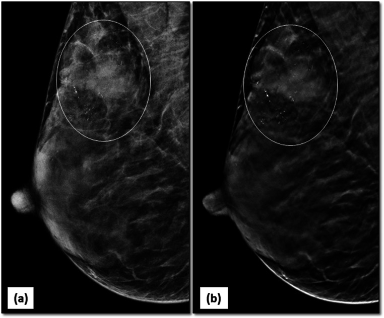
Fig. 11.
Limitation of DBT in evaluating microcalcifications: 2D MLO view (a) of right breast reveals an irregular mass with obscured margins in the posterior third of breast parenchyma in upper quadrant. Associated fine pleomorphic calcifications (circle) are seen in segmental distribution. Similar findings can be seen in the DBT image (b); however, some calcifications are accentuated however others are not well seen as the sections shows only the in-plane calcifications (circle)

