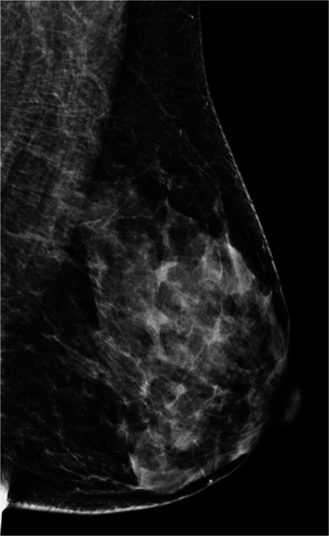
Fig. 2.

2D FFDM image in MLO view and corresponding DBT cine stack-vide (Supplementary Material) of an ACR category c (heterogeneously dense which may obscure small masses) left breast. Scrolling through the tomosynthesis stack shows normal fibroglandular parenchyma slice by slice (1 mm) without overlap, thus increasing the confidence of the reporting radiologist
