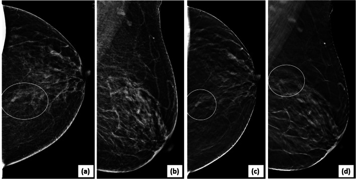
Fig. 5.
Lesion localization and characterization on DBT: a screening mammogram showed a small equal density mass (circle) with indistinct margins in the posterior third depth of left breast on 2D CC view (a). 2D MLO view (b) appeared normal. On scrolling through the CC DBT stack, the mass was best seen in the mid-slices (26/60). A targeted search in the central breast in the MLO DBT stack, revealed the lesion, as seen in selected MLO DBT slice (d). DBT (c, d) showed associated spiculations allowing accurate assessment as BI-RADS category 5 (stereotactic biopsy-invasive carcinoma)

