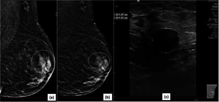
Fig. 8.
Better characterization of mass margins on DBT: 2D MLO view (a) of left breast demonstrates an oval, equal density mass (circle) in the upper quadrant of left breast with partly circumscribed and partly obscured margins (superior and inferior). The corresponding DBT image (b) delineates the previously obscured margins very well, showing them to be circumscribed (circle). The mass was designated as a BIRADS 3 lesion (likely fibroadenoma). The 6-month follow up USG image (c) of the mass confirms the circumscribed nature and stability of the mass

