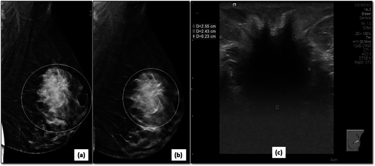
Fig. 9.
Better margin characterization in a dense breast: 2D-MLO view (a) of the left breast shows a large irregular mass (circle) in the upper quadrant of left breast. The spiculated margins (circle) are better demonstrated on DBT image (b). Corroborative ultrasound (c) confirmed the malignant features of mass as irregular, hypoechoic mass with spiculated margins and posterior shadowing. It was correctly assigned BI-RADS category 5 and biopsy confirmed an invasive ductal carcinoma

