Figure 1.

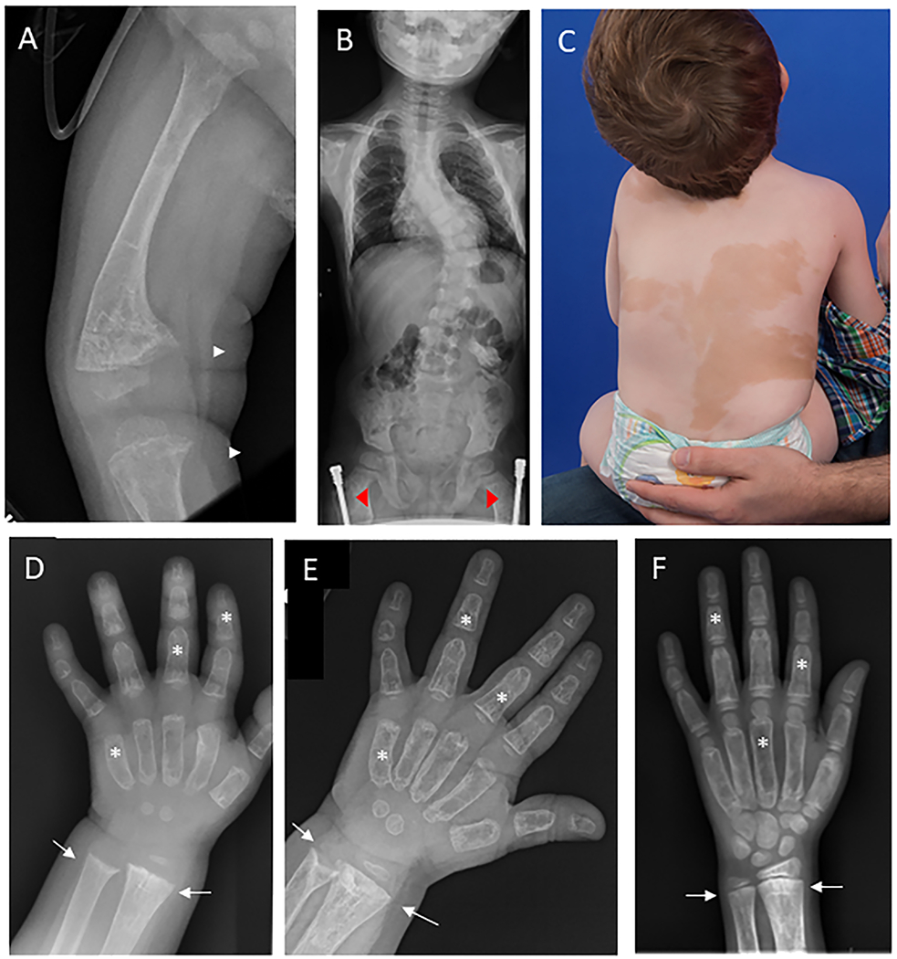
Representative clinical images. A) Radiograph at the time of hypophosphatemia diagnosis shows severe anterolateral femoral bowing and widened, frayed metaphyses at the distal femur and proximal tibia (white arrows), consistent with active rickets. The bones are diffusely involved with fibrous dysplasia, as evidenced by homogeneous “ground glass” lucency and severe cortical thinning. B) Spinal radiograph shows moderate-severe thoracolumbar scoliosis with a Cobb angle of approximately 40 degrees. Note the presence of bilateral femoral implants (red arrowheads), placed for correction of fractures and deformities. C) Photograph shows large areas of skin hyperpigmentation with features typical of McCune-Albright syndrome, including irregular borders and distribution reflecting along the midline of the body. D) hand radiograph at the time of hypophosphatemia diagnosis shows metaphyseal flaring and widening at the distal radial and ulnar metaphyses (white arrows). Note the presence of diffuse fibrous dysplasia involving all bones of the hand, leading to “ground glass” lucency and widening of the metacarpals and phalanges (white stars). E) Radiograph taken six months after starting conventional treatment demonstrates improvement in rachitic change, however there is evidence of persistent metaphyseal cupping in both the radius and ulna. F) Hand radiograph after 5 months of burosumab shows no evidence of rachitic changes at the distal radius and ulna..
