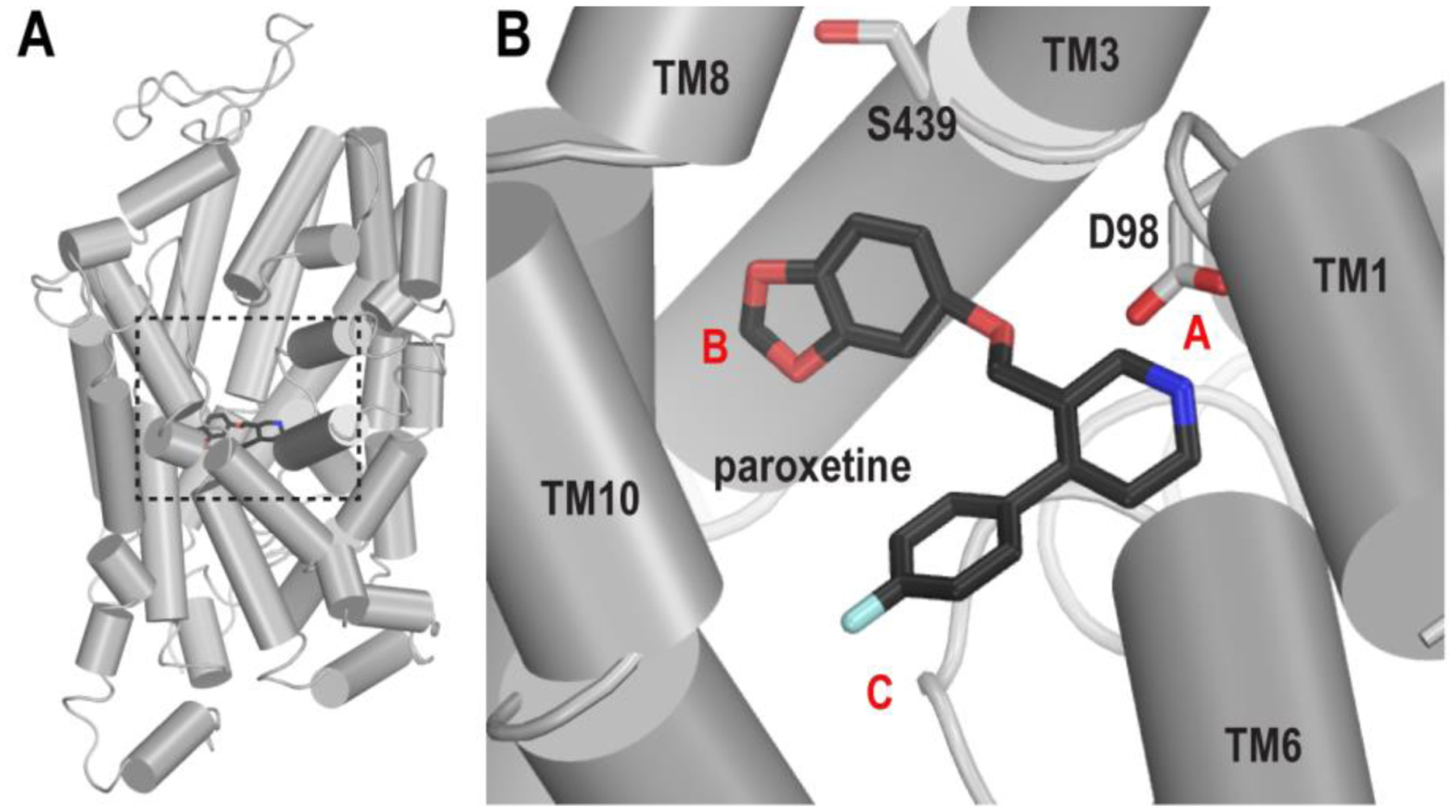
Figure 1.

The binding pose of paroxetine in the S1 site of hSERT. (A) The crystal structure of hSERT in complex with paroxetine (PDB ID 5I6X) is viewed parallel to the membrane normal in a cylindrical representation. The S1 site is enclosed by a black dotted box. (B) The paroxetine pose in the S1 site of the structure. Asp98 and Ser439 (see text) are shown in white sticks. The locations of subsites A, B, and C are labeled with red letters.
