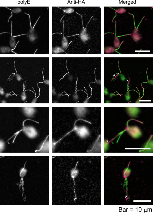
Fig. 1.
FAP70 localizes to only one microtubule bundle in frayed axonemes. fap70::FAP70-N-BCCP-HA cells were double labeled with anti-HA antibody and anti-polyE antibody, which exclusively labels DMTs. The upper row shows cells with intact axonemes; the other rows show cells with frayed axonemes. In the merged images, green is anti-polyE labeling, and magenta is anti-HA labeling. As reported previously, there is a gradient of polyE labeling of intact axonemes, with the base being more strongly labeled than the tip (upper row); this is less apparent in frayed axonemes (Kubo et al., 2010). Anti-HA labeling is more uniform; in those frayed axonemes in which it is brighter toward the tip (third row), it may reflect partial extrusion of the CA, or increased antibody accessibility to the tip region, where axonemal fraying begins. The anti-HA antibody brightly labels only one microtubule bundle in each frayed axoneme (white arrowheads).

