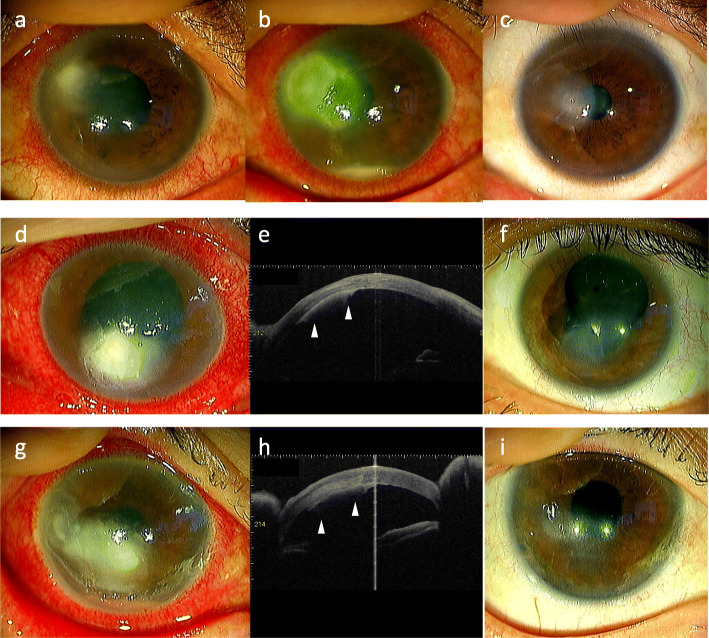
Fig. 1.
Slit-lamp and anterior segment optical coherence tomography (ASOCT) photographs obtained in each of the cases. Case #1: The feathery infiltration observed at the first visit (a) expanded to the central cornea and showed a ring-shaped lesion and hypopyon (b). At 2 months, there was keratitis scarring observed (c). Case #2: Full-thickness feathery infiltration (d) and ASOCT scanning of the retrocorneal plaque (e, arrowheads) performed at the first visit. At 2 months, keratitis scarring was observed (f). Case #3: Deep stromal feathery infiltration (g) and ASOCT scanning of the retrocorneal plaque (h, arrowheads) performed at the first visit. At 2 months, keratitis scarring was observed (i)

