
Figure 1. Spatial patterning programs that operate in the embryonic VNC and developing optic lobes.
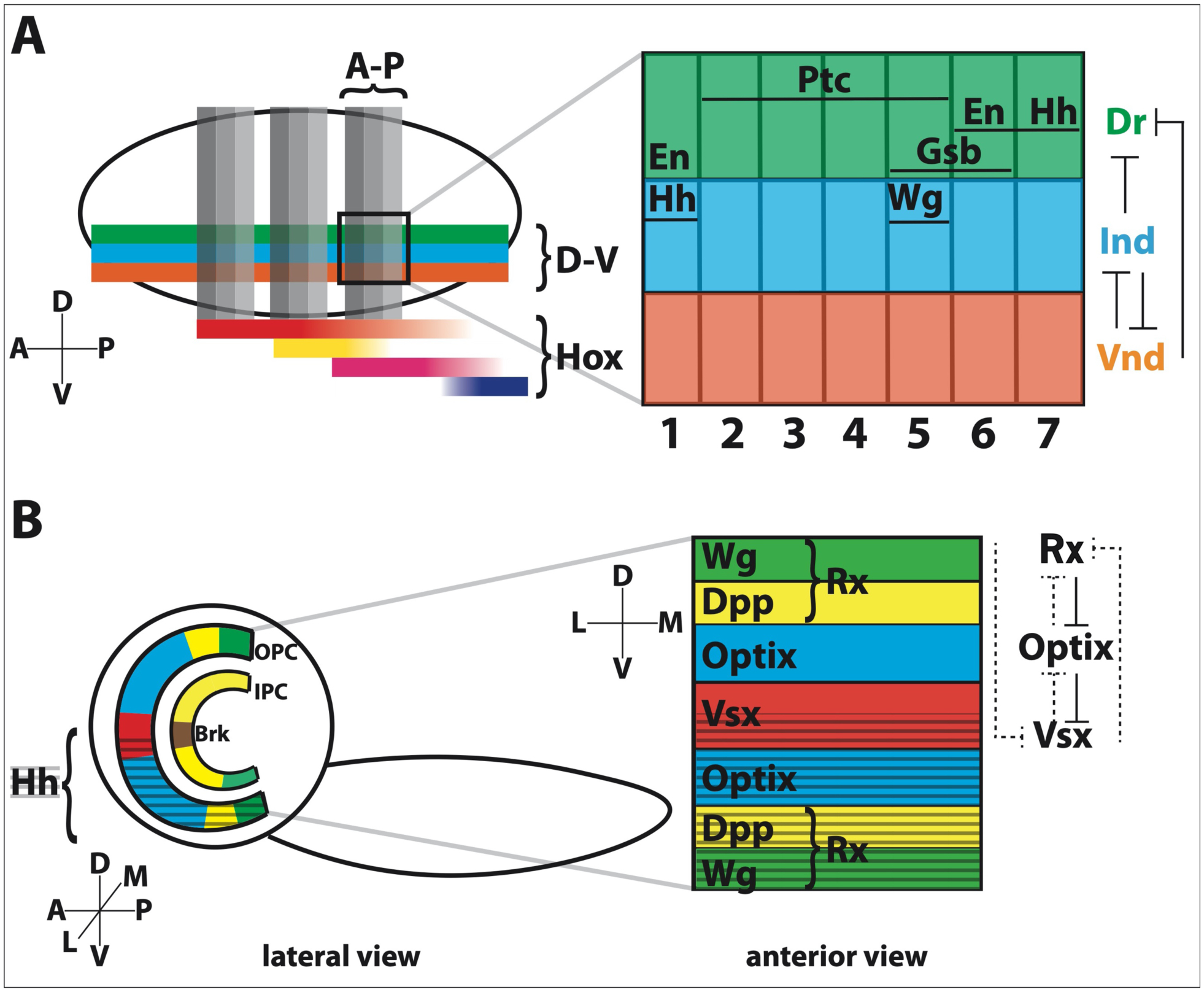
A. A simplified schematic showing how anterior-posterior (A-P) and dorsal-ventral (D-V) patterning leads to neuroblasts with unique spatial identities. Along the A-P axis, the VNC is subdivided into 14 repeated segments, represented here by vertical gray bars. Within each segment, the segment-polarity genes define different rows (numbered 1–7). The D-V axis is subdivided into three columns, represented here by green, blue, and orange bars. Each segment is further defined by the expression of different Hox genes. One segment is magnified to show the expression domains of the different sTFs in each array. Only the interactions between the D-V sTFs are shown for simplicity. En: Engrailed; Hh: Hedgehog; Ptc: Patched; Wg: Wingless; Gsb: Gooseberry; Dr: Drop (also called Msh); Ind: Intermediate neuroblasts defective; Vnd: Ventral nervous system defective.
B. A simplified larval brain is viewed from the side (lateral) to show the crescent shape of the OPC and IPC, which give rise to optic lobe neuroblasts. The OPC can be subdivided into 8 compartments and the IPC into 4 compartments along the D-V axis. The ventral hedgehog (hh) domain (marked by gray hashed lines) is a remnant of earlier developmental expression. Unlike the VNC, there is no indication that the OPC or IPC are spatially patterned along a second axis (i.e., the medial-lateral axis representing the posterior-anterior retina), which is illustrated by rotating the view of the OPC by 90 degrees (clockwise). The boundaries defining D-V compartments are maintained by interactions between the sTFs themselves.