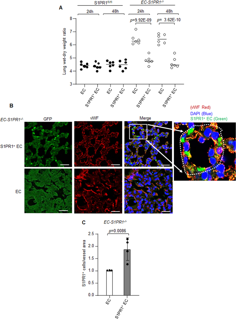
Figure 7: S1PR1+ endothelial cells promote resolution of lung endothelial injury.
A, S1PR1+ EC (GFP+CD31+CD45−) or non-GFP EC (GFP−CD31+CD45−) were flow sorted at 16h post LPS challenge (10 mg/kg i.p.) of S1PR1-GFP reporter mice. S1PR1+ EC or EC (~1.0×10⁶) were then injected i.v. into S1PRfl/fl or EC-S1PR1 null mice. Subsequently, lung vascular injury was determined by measuring lung wet-dry weight ratio after 24h or 48h post transplantation (n=6 mice/group). B, a micrograph from EC-S1PR1 null lungs receiving S1PR1+ EC or non-GFP EC were stained with anti-GFP and anti-vWF antibodies to assess integration of S1PR1+ EC into intima of the vessel. Micrograph of experiments were repeated multiple times. Scale bar 50 μm. C, respective quantification of S1PR1+ EC quantified as number of S1PR1+ EC over vessel area (n=4). A and C show individual data along with mean ±SD. Data in A were analyzed using one-way ANOVA followed by Tukey’s multiple comparisons test, whereas unpaired t test was used for C (See also Online Table II). A, p=9.92E-09, p= 3.62E-10 indicate significance relative to mice receiving EC and C, p=0.0086 indicate significance relative to EC.

