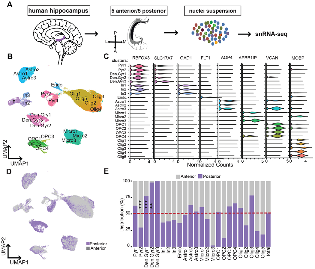
Figure 1. Unbiased snRNA-seq analysis identified 24 distinct cell types in human anterior and posterior hippocampus samples.

(A) Schematic overview of the experimental procedures used to extract nuclei from 5 anterior and 5 posterior samples, single-nuclei capture and barcoding using 10X Genomics Chromium, and Illumina next-generation sequencing. (B) UMAP plot of all cells analyzed from anterior (64,076) and posterior (65,832) hippocampus, colored by cluster identities and cell-type annotations. Pyr=Pyramidal neurons, Den.Gyr=dentate gyrus neurons, In=Interneurons, Endo=endothelial cells, Micro=microglia, Astro=astrocytes, OPCs=oligodendrocyte progenitor cells, and Olig=oligodendrocytes. (C) Violin plots of expression values for cell-type-specific marker genes. (D) UMAP plot of all cells analyzed, colored by the axis the cells were recovered from (posterior, purple; anterior, gray). (E) Bar chart showing the frequency distribution of all clusters between posterior (purple) and anterior (gray). *P < 0.01, ***P < 0.001, Robust generalized mixed model. See also Figure S2, S3, S4, and S5 and Table S2, S3.
