
Figure 3. In vitro anticancer efficacy against OSI-resistant PC-9/AR NSCLC cells.
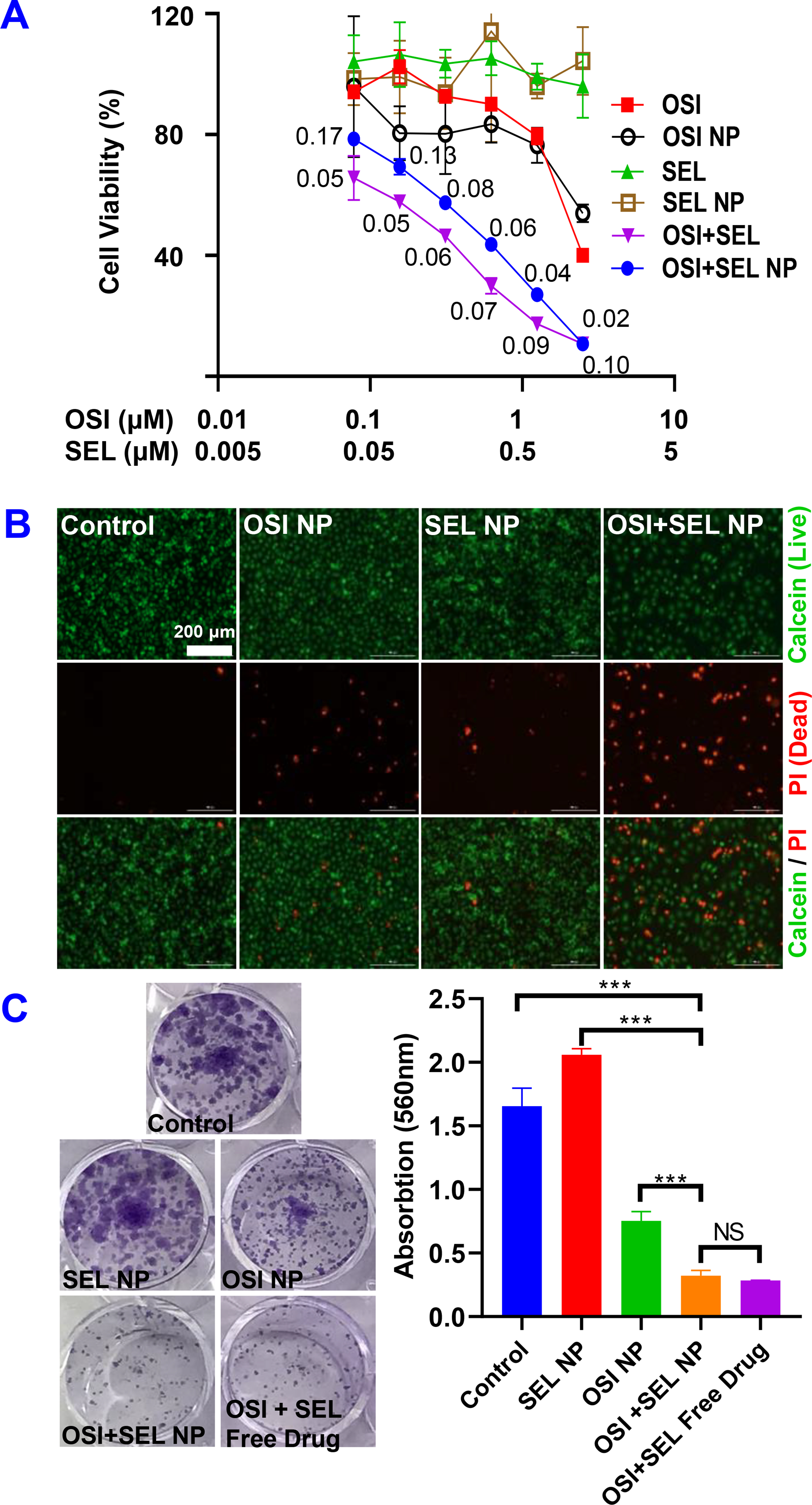
(A) Cell viability of PC-9/AR cells were determined with the MTT assay after treatment with different formulations for 48 hours. The numbers in the graphs are combination indices. (B) Calcein-AM/PI staining. Cells treated with different formulations for 48 hours and stained with Calcein-AM/PI to detect living (green) and dead (red) cells. (C) Inhibition of colony formation by OSI+SEL combination therapy. Cells were treated with different NP formulations and free drugs for a total of 7 days. Cell colonies were stained with crystal violet and photographed. Crystal violet in each groups were also dissolved by methanol and the absorption at 560 nm were determined. Results are mean ± SD (n=3). ***, P < 0.001