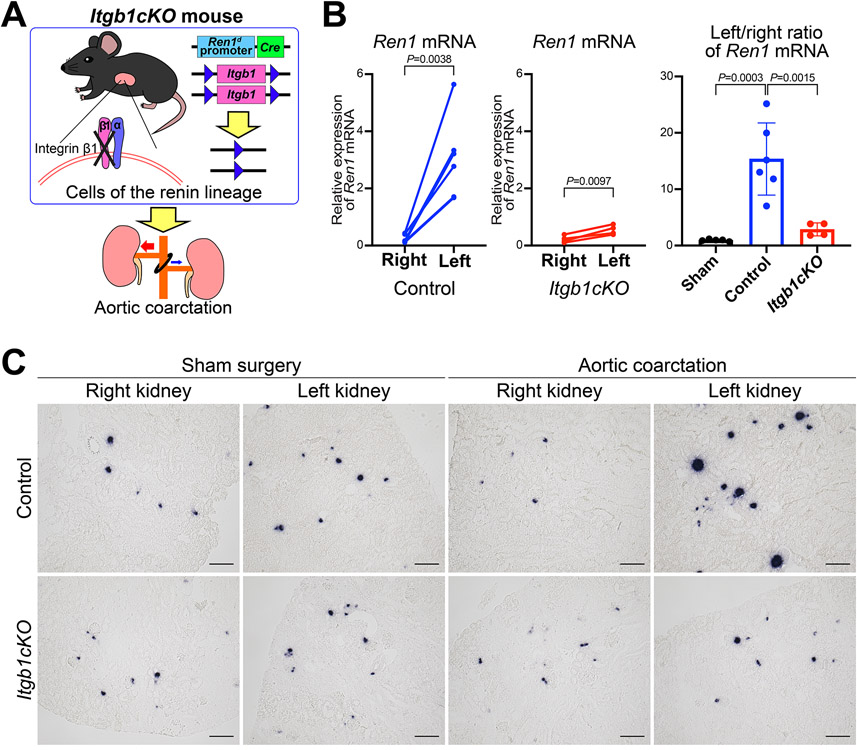
Figure 4. Itgb1 gene knockout in cells of the renin lineage inhibited the response to changes in perfusion pressure.
A, Mice with conditional deletion of the Itgb1 gene in cells of the renin lineage (Itgb1cKO) and control mice were subjected to aortic coarctation (AoCo). B, Itgb1cKO mice showed impaired responses in Ren1 expression to changes in perfusion pressure. Quantitative reverse transcription PCR showed significant differences in Ren1 mRNA between the right and left renal cortices from the control mice (n=6, paired t-test) and from the Itgb1cKO mice (n=4, paired t-test), respectively. The left/right ratio of Ren1 mRNA of Itgb1cKO mice was significantly lower than the one of control animals (one-way ANOVA followed by Tukey’s multiple comparison test). C, in situ hybridization for Ren1 mRNA in the kidneys from Itgb1cKO mice subjected to the AoCo. For Itgb1cKO mice, the decrease in Ren1 mRNA in the right kidneys and the increase in that in the left kidneys were milder than those in control mice, respectively. Scale bar, 100 μm. All data are reported as means ± standard deviation. **P<0.01, ***P<0.001.

