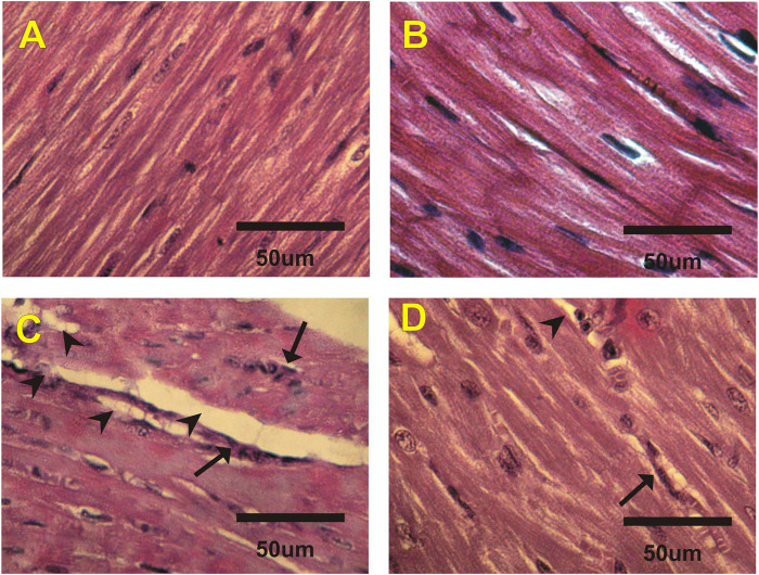
FIGURE 2.
Photomicrographs of heart sections from different experimental groups stained with H&E. The (A) control (G1) and (B) costus (G2) groups show a normal myofibrillar structure with striations. (C) The EST group (G3) shows moderate myocardial atrophy and leukocyte infiltration (arrows). (D) The EST group treated with costus (G4) shows mild myocardial atrophy. H and E, hematoxylin and eosin; EST, Ehrlich solid tumor.

