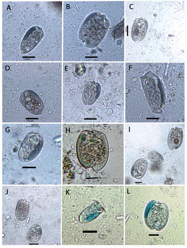
Figure 2.
Photomicrographs of some entodiniid ciliates observed in the rumen of European bison. (A) Entodinium brevispinum; (B) E. caudatum; (C) E. lobosospinosum; (D) E. simplex; (E) E. nanellum; (F) E. rostratum; (G) E. parvum; (H) E. yunense; (I) (from left to right) E. simplex, E. nanellum, and E. yunense; (J) E. orbicularis; (K) E. exiguum; and (L) E. dubardi. Samples were colorized by (A–J) chrome-alum-carmine and (K,L) methyl-green formalin solutions. Scale bars are 10 μm in all pictures.

