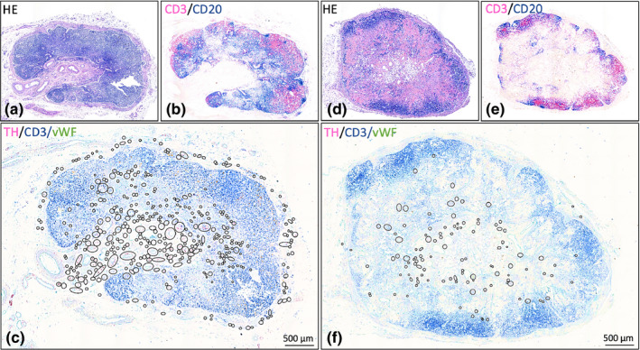
FIGURE 1.

Lymph nodes with different quantities of sympathetic nerves. Hematoxylin / eosin and CD3/CD20 stained slides show the general lymph node morphology and distribution of lymphocytes, respectively. In overview images of TH/CD3/vWF stained slides, the presence of sympathetic nerves in the various compartments is indicated by black circles (in these overview images, nerves cannot be distinguished). More detailed images of sympathetic nerve distribution can be found in Figure 2. (a‐c): Lymph node (# 13) with high nerve quantity. (d‐f): Lymph node (# 15) with low nerve quantity. Black circled structures: sympathetic nerves ( Small circles: discrete nerve fibers or small nerves Larger circles: clusters of nerves / blood vessels with paravascular nerves)
