
FIGURE 2.
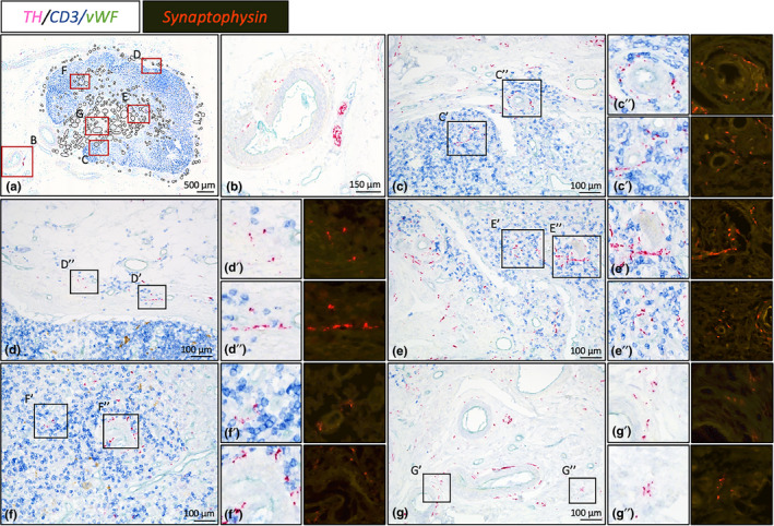
Magnified images of sympathetic innervation of lymph node # 13. Bright field images represent TH/CD3/vWF triple stained slides showing sympathetic nerves (in relation with), T cells and blood vessels, respectively. Synaptophysin stained slides show the presence of varicosities. (a): Overview image of lymph node #13. Black circled structures represent locations with sympathetic nerves. Red boxed areas are shown in more detail in figures B‐G. (b): Large sympathetic nerve bundle running with a lymph node artery towards the hilum. (c‐g): Nerves can be observed as paravascular or as discrete entities of which some are in close proximity with T cells. (c): Cortex, (d): Capsule, (e): Medulla, (f): Cortex, (g): Hilum