
FIGURE 2.
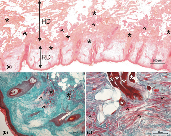
(a) Overview of the skin region next to the orbicularis oris muscle. Note the numerous muscle fiber bundles (asterisks) and sweat gland formations (arrowheads) in the hypodermis (HD). The reticular dermis (RD) appears pale. The thin papillar dermis follows the hair follicles. (b) Muscle fibers enter the dermis (asterisks) and end in regions with high amounts of elastic fibers (arrowheads). (c) Higher magnification shows the brush‐like arranged protrusions of the muscle fibers (asterisks); the elastic fibers (red, marked by arrowheads) are not parallel to the muscle fibers; in addition, the collagen fibers (green) are not densified in the regions of muscle fiber endings (a: HE staining; b,c: Goldner staining)