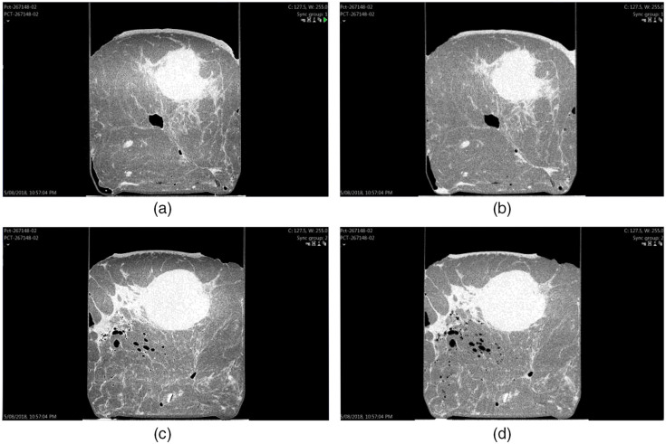
Fig. 1.
Screen setup of PB-CT versus AB-CT images. This case was a post neoadjuvant chemotherapy of invasive carcinoma, no special type, no residual in-situ, or invasive disease (sample ID: 267148); (a) axial PB-CT slice; (b) axial AB-CT slice; (c) sagittal PB-CT slice; (d) sagittal AB-CT slice; PB-CT images for this image set were scanned at an x-ray energy of 28 keV; AB-CT images were scanned at an x-ray energy of 32 keV.

